
Outline EEG Overview Purpose Indications Type of EEG Tests Nursing Interventions Patient Preparation Patient and Family Teaching Normal Abnormal Results ID: 644700
Download Presentation The PPT/PDF document "Electroencephalogram EEG" is the property of its rightful owner. Permission is granted to download and print the materials on this web site for personal, non-commercial use only, and to display it on your personal computer provided you do not modify the materials and that you retain all copyright notices contained in the materials. By downloading content from our website, you accept the terms of this agreement.


Slide1
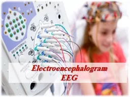
Electroencephalogram
EEG Slide2
Outline;
EEG Overview.
Purpose
Indications
Type of EEG Tests
Nursing Interventions;
*
Patient Preparation.
*
Patient and Family Teaching.
Normal / Abnormal Results
Common Factors affecting EEG Recording
Complications.Slide3
Objectives
At the end of the lecture, the students will be able to
Understand EEG procedure.
Discuss nursing management.Slide4
Diagnostic Tests for Neurological Diseases
Brain scans
Cerobrospinal
fluid analysis
Computed Tomography (CT scan)
,
Echocardiogram
Electroencephalography (EEG)Slide5
The Electroencephalogram (EEG
)
Definition:
Is a medical test used to measure the electrical activity of the brain, via electrodes applied to scalp
or through microelectrodes placed within the brain tissueSlide6
Electrodes
are attached to multiple sites on the scalp to provide a recording of electrical activity that is generated in the cerebral cortex.
Electrical impulses
are transmitted to an electroencephalograph, which magnifies and records these impulses as brain waves on a strip of paper.Slide7
PURPOSE
It
provides a physiologic assessment of cerebral activity.
for
diagnosing and evaluating seizure
disorders, epilepsy,
coma, or organic brain
syndrome, sleep disorders.
Used in making
a determination of brain
deathused
in intensive care units for brain function monitoring non-convulsive seizures/non-convulsive status
epilepticus
effect
of sedative/anesthesia in patients in medically induced coma (for treatment of refractory seizures or increased intracranial
pressure) secondary brain damage in conditions such as subarachnoid hemorrhage Slide8
Where is it performed?
room with no electrical interference;
bedside
-
comatose patient
(
using a portable unit.)
The
test takes about 1 to 2 hour.
completely painless , no need for shaving the hair.
Provides
information about the timing of events.
Indication:
epilepsy,
coma,
sleep disorders,
confirmation of brain death
Tumors, brain abscesses, blood clots, and infection may cause abnormal patterns in electrical activity. Slide9
For a baseline recording
,
the patient
is instructed to
lie
quietly with both eyes closed
(
resting phase)
hyperventilate for 3 to 4 minutes and then look at a bright, flashing light for stimulation.
( activation procedures)
to evoke abnormal electrical discharges, such as seizure potentials. Slide10
During the recording, a series of activation procedures may be used. These procedures may induce normal or abnormal EEG activity that might not otherwise be
seen. (hyperventilation
, photic stimulation (with a strobe light), eye closure, mental activity, sleep and sleep
deprivation).
A sleep EEG
may be recorded after sedation because some abnormal brain waves are seen only when the patient is asleep. Slide11
Types of EEG test
:
Routine EEG tests -
EEG test done
at an outpatient’s appointment at the
hospital (
lasts about one
hour)
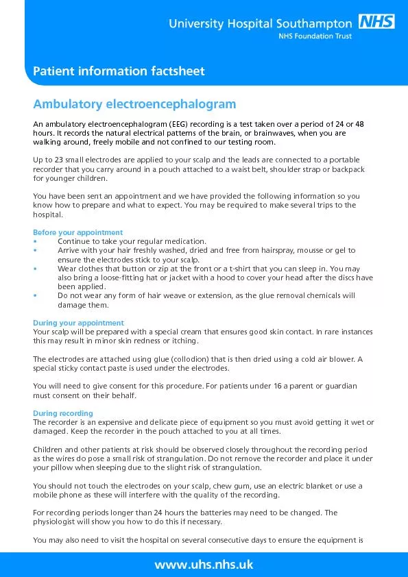
2. Ambulatory
EEG
test- recording the activity in the brain over a few hours, days or weeks, allowing
more time for the test to pick up any unusual electrical activity in the brain, the electrodes are plugged in to a small monitor that records the
results.
3. Sleep
EEG
tests - an EEG test is done while the patient is asleep, usually done in hospital, using a routine EEG machine. Before the test, the patient may be given some medicine to induce sleep. The test lasts for one to two hours or up to 8 hours Patient goes home once he wakes up 4. Sleep-deprived EEG tests - done when a patient had less sleep than usual. Before a sleep-deprived EEG test, the patient is advised not to go to sleep at all the night before, or just to wake up much earlier than usual. A patient tries to fall asleep or doze while the EEG is still recording the activity in the brain. The test lasts for a few hours. Patient goes home once he wakes upSlide12
Types of EEG test
:
5. Video-telemetry tests – a patient wears an ambulatory
EEG usually
carried out over a few days
. All
movements are recorded by a video camera.
The patient
need to stay in
hospital
This is done if a patient have already been diagnosed with epilepsy to determine the following:
To determine the type of seizuresThe reason / cause why anti-epileptic drugs are not working
well
Other
possibility that
the seizures are caused by other etiology than epilepsy.considering having epilepsy surgery.Slide13
Nursing Interventions
A- Patient Preparation;
Explain the procedure to the patients, emphasizing the importance of cooperation.
Withhold
Antiseizure
agents, tranquilizers, stimulants, and depressants medications for 24 to 48 hours before an EEG (alters the EEG wave patterns or mask the abnormal wave patterns of seizure disorders)
NO coffee, tea, chocolate, and cola drinks in the meal before the test (stimulants)Slide14
Have regular meal before the EEG
( to avoid alteration of blood glucose level. Low blood sugar can cause changes in the brain wave patterns and change the EEG result)
Assure the patient that the procedure does not cause an electric shock and that the EEG is a diagnostic test, not a form of treatment.
Patients with seizures do not stop taking their
antiseizure
medication prior to testing.
Assist the patient to wash the hair before and after the test.Slide15
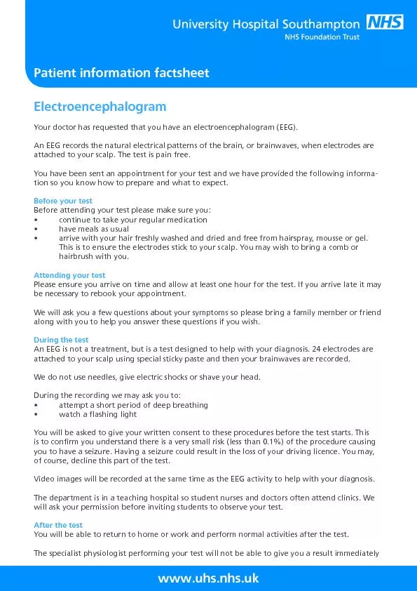
B- Patient and Family Teaching
The test takes about 1 to 2 hour.
The test is painless and will be performed while sitting in a comfortable chair or lying on a stretcher.
The electrodes are applied to the scalp with a thick paste.
During the test, the patient will first be asked to breathe in and out deeply for a few minutes. Then, close her eyes while a light is flashed on them and, finally, the patient will lie quietly with her eyes closed.
After the test, the nurse will help the patient wash the paste out of the patient’s hair.Slide16
Normal Results
Brain
electrical activity has certain frequencies (the number of waves per second) that are normal for different levels of consciousness. For example, brain waves are faster when the person is awake
, and slower when
he/she is
sleeping.
There
are also normal patterns to these waves.
The
frequencies and patterns are what the EEG reader looks for.
Note: A normal EEG does not mean that a seizure did not occurSlide17
Abnormal
Results
Abnormal results on an EEG test may be due to:• An abnormal structure in the brain (such as a brain tumor)
• Attention problems
• Tissue death due to a blockage in blood flow (cerebral infarction)
• Drug or alcohol abuse
• Head injury
• Inflammation of the brain (encephalitis)
• Hemorrhage (abnormal bleeding caused by a ruptured blood vessel)
• Migraines (in some cases)
• Seizure disorder (such as epilepsy or convulsions)• Sleep disorder (such as narcolepsy)Slide18
Common factors affecting EEG recording ( artifacts
)
The biggest challenge with monitoring EEG is artifact recognition and elimination.
There
are patient related artifacts (e.g. movement, sweating, ECG, eye movements
)
technical artifacts
(cable
movements, electrode paste-related), which have to be handled differently.
Common
factors are:• Radio frequency (RF) waves• Electromagnetic interference • Eye movement
• ECG
• Head movement
• Muscle
• Sweating• Electrode Slide19
Complications
EEG is a safe test with
no side effects.
However, a person with epilepsy may experience a seizure, triggered by the various stimuli used in the procedure, including the flashing lights.
This is not seen as a 'complication' by medical staff, because a seizure during an EEG can greatly help in diagnosis.)Slide20