

skiffy slipped upper femoral epiphysis Done by Yara Saleh outlines 1 Definition 2Epidemiology 3Presentation 4Clinical features 5Diagnosis 6Complications 7Treatment normally a growing femur has 4 main parts ID: 917333
Download Presentation The PPT/PDF document "Slipped capital femoral epiphysis (SCFE ..." is the property of its rightful owner. Permission is granted to download and print the materials on this web site for personal, non-commercial use only, and to display it on your personal computer provided you do not modify the materials and that you retain all copyright notices contained in the materials. By downloading content from our website, you accept the terms of this agreement.


Slide1
Slipped capital femoral epiphysis (SCFE or skiffy, slipped upper femoral epiphysis)
Done by :
Yara
Saleh
Slide2outlines1- Definition 2-Epidemiology 3-Presentation
4-Clinical features
5-Diagnosis
6-Complications
7-Treatment
Slide3*normally a growing femur has 4 main parts :1- diaphysis 2-metaphysis
3- growth plate also called
physis
4-epiphysis *The cartilaginous growth plate has cells that divide and enable the bone to grow in length , these cells are very active in adolescents and they enable a growth spurt*During this period a growth plate is very weak and vulnerable to shearing force*Before the growth plate is ossifies it supported by perichondrial ring which is CT that extends from metaphysis to the epiphysis , it helps resist the shearing forces so the femoral head and neck don’t slip away from each other * Eventually the cartilaginous growth plate ossifies and fuse with epiphysis
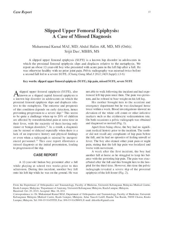
Slide4DefinitionSCFE is the most common hip disorder in adolescence.SCEF is a medical term referring to a fracture through the growth plate (
physis
), which results in slippage of the overlying end of the femur (epiphysis) from the femoral neck caused by weakness of the
perichondral
ring.The femoral epiphysis remains in the acetabulum, while the metaphysis move in an anterior direction with external rotation.
Slide5Slide6EpidemiologyBoys are affected more often than girls.
SCFEs often occur in :
1-In obese children
2-males (male to female ratio is 2:1.4)3-during period of rapid growth (10-16 years of age)Two-thirds of the patients are overweight and sexually under-developed, or unusually tall and thin.If one side slips there is a 30% risk of the other side slipping as well.
Slide7Presentation*Chronic (>3 weeks): most common
Aching groin, hip, thigh or knee pain and limp
- Often no history of trauma
*Acute (<3 weeks):
severe hip pain, inability to walk usually after trauma *Minor vs moderate vs. severe depends on displacement of epiphysis in relation to the diameter of the femoral neck.-Minor displacement: displacement of less than one-third the width of the epiphysis-Moderate displacement: displacements of one third to one-half the epiphyseal width- Severe displacement: if the displacement is more than one-half the epiphyseal width
Slide8Clinical features The patient presents with gradual, progressive onset of pain in the groin, the anterior part of the thigh or the knee (referred pain); he may also limp.
On examination the leg is externally rotated and is 1 or 2 cm short. Characteristically there is limitation of abduction and medial (internal) rotation. Following an acute slip, the hip is irritable and all movements are accompanied by pain.
Slide9Diagnosis By pelvic Xray
*AP radiograph:
Klein line is a line drawn along the superior border of the femoral neck that would normally pass through a portion of the femoral head. If not, slipped capital femoral epiphysis is diagnosed. →
Trethowan's
sign
Slide10Slide11* Frog leg radiograph: Is diagnostically more reliable; even minor degrees of slip can be shown by drawing lines through the base of the epiphysis and up the middle of the femoral neck – if the angle indicated is less than 90 degrees, the epiphysis has slipped
posteriorly
.
-note :
A straight line through the center of the femoral neck proximally should be at the center of the epiphysis. If not, and the line is anterior in the epiphysis, it is likely an SCFE.
Slide12Slide13Complications1-Slipping at the opposite hip
occurs in one-third
of cases.
2-Avascular necrosis is the most
serious complication.3-Coxa vara deformity may result if the displacement is not reduced and the epiphysis fuses in its deformed position. The patient limps but the condition is usually painless. Osteotomy may be needed to correct the deformity.4- Secondary osteoarthritis is a likely sequel if displacement has not been reduced, and inevitable if there has been avascular necrosis.
Slide14Treatment - Surgery, either In situ fixation using screws or osteotomy if there's deformity.
- Closed reduction is dangerous and should not be attempted
Slide15Thank you