
b y Krisztina HMinkó Semmelweis University Faculty of Medicine Department of Anatomy Histology and Embyology T he initially flat threelayered embryonic disc undergoes ID: 998685
Download Presentation The PPT/PDF document "TERATOLOGY for pharmacist" is the property of its rightful owner. Permission is granted to download and print the materials on this web site for personal, non-commercial use only, and to display it on your personal computer provided you do not modify the materials and that you retain all copyright notices contained in the materials. By downloading content from our website, you accept the terms of this agreement.


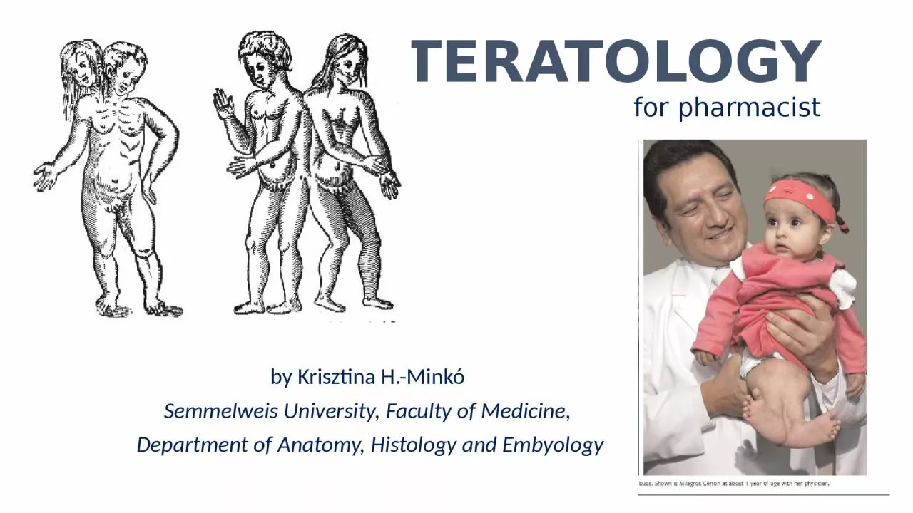
1. TERATOLOGYfor pharmacistby Krisztina H.-Minkó Semmelweis University, Faculty of Medicine, Department of Anatomy, Histology and Embyology
2. The initially flat three-layered embryonic disc undergoes morphogenesis to form a three-dimensional embryo with a tube-within-a-tube body plan and the beginnings of rudiments that will form all of the adult organs andsystems.Morphogenesis results from differential growth. Differential growth is driven by a small number of fundamental cellular behaviors such as changes in cell shape, size, position,number, and adhesivity. If these behaviors are perturbed duringembryogenesis, by a genetic mutation, environmentalinsult (i.e., a teratogen), or a combination of the two, differential growth is abnormal and dysmorphogenesisresults with the formation of a structural birth defect.
3. Dysmorphogenesis can result from both malformationand deformation. Malformations consist of primary morphologic defects in an organ or body part resulting from abnormal developmental events that are directly involved in the development of that organ or body part. For example, failure of the neural groove to close results in a malformation called a neural tube defect. Similarly, failure of the digits to fully separate results in syndactyly, that is, fusion of the digits.Deformations consist of secondary morphologic defects that are imposed upon an organ or body part owing to mechanical forces; that is, deformations affect thedevelopment of an organ or body part indirectly. Forexample, if insufficient amniotic fluid forms (i.e., oligohydramnios), deformation of the feet can occur dueto mechanical constraints, resulting in club foot.Dysmorphogenesis can occur in an isolated organ orbody part or can occur as a pattern of multiple primarymalformations with a single cause. In the latter case, thecondition is referred to as a syndrome. Common examples,include Down syndrome (trisomy 21) and 22q11.2 deletion syndrome, two syndromes that result from genetic mutations.Other syndromes can result from teratogen exposure.A common example is fetal alcohol syndrome,also known as fetal alcohol spectrum disorder.
4. Teratogens are environmental (i.e., nongenetic) substancesthat are capable of causing a birth defect whenembryos or fetuses are exposed at critical times indevelopment to sufficiently high doses (concentrations).The study of the role of environmental factorsin disrupting development is known by the unfortunatename of teratology, which literally means thestudy of (developmental) monsters.
5.
6. CONGENITAL MALFORMATION, TERATOLOGY, HISTORYTeratology is the science that studies the causes, mechanisms, and patterns of abnormal development. teratology : development of „monsters” 1941 Gregg (Australia) rubella infection of pregnants caused blindness, deafness , problems with heart1960s years: Thalidomide scandal (phocomelia)
7. The first principle of teratology is that an embryonic structure is usually susceptible to teratogens only during specific critical sensitive periods, which usually correspond to periods of active differentiation and morphogenesis.Thus, a potent teratogen may have no effect on the development of an embryonic structure if it is administered before or after the critical period during which that structureis susceptible to its action.Because the major events of organogenesis take place during the first 8 weeks of development, that is the period during which the fetus is most vulnerable to teratogens.
8. A second principle of teratology is that an embryonic structure is susceptibleto a critical dose of teratogen during its specific criticalsensitive period. Thus, in teratologic studies a dose responsecurve is constructed for a suspected teratogenin which lowest dose has no effect and the highestdose is lethal to the embryo.A third principle of teratologyis that susceptibility to a teratogen dependson the genetic constitution of the developing embryoor fetus. For example, if two embryos of the same ageare exposed to the same dose of teratogen, one maydevelop severe cardiac malformations whereas theother may remain unaffected. The molecular basisfor this difference in susceptibility might, for instance,be a genetic difference in the rate at which the enzyme systems of the two embryos detoxify the teratogen.Thus, there is a gene-environmental interactionunderlying susceptibility to birth defects that variesfrom embryo to embryo.
9. Incidence of abnormalities in Europe: 2-3% of all live born children at the birth,-(congenital)but in the first year: 4-6%.Range of CONGENITAL ANOMALIES is very videfrom the severe morfological defects till enzime defects (no morfological picture)
10. Developmental abnormalitiesgenetic sourcesEnvitonmental sources(teratogens) CAUSES OF CONGENITAL ANOMALIES
11. unknown 50%Chromosome mutation 10%Gene mutation 8%Umwelt 7%multifactorial 25% ??:- age of the parents different ethnical groups familiar background (genetical) seasons social and geographical differences
12. Incidence of Down syndrome is dependent on the age of mother (left figure) but the Apert syndrome is on the age of father (right blue line)Apert syndrome: turricephalus – tower headAGE
13. Incidence of cleft palate is different in different ethnical groupsfar-east > (2x) caucasian > (2x) afro-americanRACE
14. Anencephaly is abundant in the spring: it is caused by the deficit of the mother’s folic acid level. Given folic acid to the mothers cut down drastically the number of these fetuses.Anencephaly = cranioschisis – anterior part of the neural tube (neuroporus anterior) is not closed the brain will not be developed.SEASON
15. Frequency of anencephaly in different countries
16. Critical periods during the pregnancyPre-embryonic period:0-3. embrionic weeks (EW): „to be or not to be”, the teratogen kills the whole embryo (abortion), or only some of the early cells and these cells will be substituted (no congen. malf.)Embryonic peroid:3-8. EW: most vulnerable period: time of the development of the most serious (major) anomalies Fetal period:8. EW: minor morphological or functional defects (e.g.mental retardation).
17. The sensitivity of the different organs are different in the time: some organs are sensitive in early period (heart) the others are sensitive in later periods (genitals) and some of them are sensitive all along (nervous system).Diagram of sensitivity period
18.
19. Different organs different sensitive periods Tetraciklin: - after the 120. embrionic day (ED) : milk-teeth and adultteeth become coloured-after ED250: only the adult teeth become colouredRubella infection:ED0 - ED60: cataracta (blindness )and heart problemsED0 - ED120: deafnesshttps://drmarthaszabolcs.com/2017/03/06/first-blog-post/#jp-carousel-140
20. Causes of CAgenetic factorschromosome abnormalitiesNon-disjunction abnormal number of cromosome: aneuploidia monosomy trisomyabnormalities inside the chromosome reciprocal translocation deletion,duplication
21. Aneuploidia of the autosome chromosomes:Down syndrome: 21 trisomiasymptomes: defects of the face, mental retardation, typical crease on the palm>35 age at pregnancy – higher riskWhy these signs result from the trisomy is unknown
22. Aneuploidia of the autosome chromosomes:Down syndrome: 21 trisomiasymptomes: defects of the face, mental retardation, typical crease on the palm>35 age at pregnancy – higher riskWhy these signs result from the trisomy is unknown
23. Other trisomies:Trisomy of 13. Trisomy of 18. (rocker bottom feet)
24. Aneuploidia of the sex chromosomes:Turner syndrome: 45 X,O (X monosomy): female but weak fenotype, sterilOthers:XXY: Klienefelter syndrome: male, but small testis, steril, long limbsXYY: seems to be normal male, high figure, but violent (choleric) behaviorXXX: „super women”: seems to be normal feminin habit, but mental retardation is occure
25. abnormalities inside the chromosome reciprocal translocation deletion, duplicationcause specific symptomes e.g. :
26. Androgen insensitivity syndromeHere it is a niceman !!!!!!(he has testis)but: NO prostate, and any other male genital organs!NO ovary and female genital organs, only blind vaginaIt’s not rarely at photomodells...…
27. Andrej Pejic, the gender-fungible Australian male model
28. Testicular feminizationIn this case the genotype is normal male ( XY) , the first steps of the sex determination is normal : the testis is developed, but the testosteron produced by the testis not able to act on the cells because the testosteron receptors are missing on the cells
29. Syndaktilia: az ujjak összeolvadása. Ebben a humán esetben a HOXD13 gén pontmutációja a gén kieséséhez vezetett. Az üres csillagokkal jelölt képletek kézközépcsontok, a két kicsi csillag normálisan nem létező carpalis csontokat jelöl.HoxD13 pointmutation (polysyndactylia)
30. Holt-Oram syndrome:-TBX5 mutation-1:100.000-limb bud (100%)phocomelia - heart defects (67%)
31. Causes of CAEnvironmental factors 1. biotic agents (bacteria, viruses, fungi) 2. drugs/chemicals 3. mechanical agents
32. 1.Infectious Agents TeratogensRubella German or Three-Day MeaslesFirst trimester – most serious ED0 - ED60: cataracta (blindness )and heart problemsED0 - ED120: deafnessCytomegalovirus (CMV)Herpes Simplex Virus (HSV)VaricellaHuman Immunodeficiency Virus (HIV)Congenital SyphilisToxoplasmosis
33. 2. Mechanical Factors as TeratogensMechanical forcesRestrict the mobility of the fetusCause prolonged compression in an abnormal postureE.g. congenital dislocation of hip and clubfootMalformed uterus
34. Mechanical Factors as TeratogensAmniotic fluidAbsorb mechanical forcesOligohydramniosSignificantly reduced fluid-quantityMechanically induced deformations of the limbsKnee hyper-extendsAmniotic bandsRings formed from amnion ruptureLocal constriction during fetal growthIntrauterine amputations
35. 3. DRUGS/CHEMICALSEtil-alcohol: Fetale Alcohol Syndrome (FAS): causes problems in the brain development : holoprosencephalia. Prosencephalon is the most anterior part of the neural tube : hemispheres, hypothalamus, hypophysis , eye developed from this partThalidomid (Contergan): tranquillizierMany therapeutic drugs are known to be teratogenic;these include retinoids (vitamin A and analogs), theanticoagulant warfarin, the anticonvulsants valproicacid and phenytoin, and a number of chemotherapeuticagents used to treat cancer. Most teratogenic drugsexert their main effects during the embryonic period.Although, as stated above, most care must be exercisedin administering certain anesthetics and other drugseven late in pregnancy or at term, because they mayendanger the health of the fetus.
36. FAS: holoprosencephaly and craniofacial CA.Prosencephalon regulate not only the development of the brain but the head and face alsoB: cebocephaliaC: no nose, hypotelorismusD: cyclopia
37. Some recreational drugs are also teratogenic; theseinclude tobacco, alcohol, and cocaine. Cocaine, used by alarming numbers ofpregnant women (the drug affected 300,000 to400,000 newborns in 1990 in the United States), readilycrosses the placenta and may cause addiction in thedeveloping fetus. In some of the major cities of theUnited States, as many as 20% of babies are born tomothers who abuse cocaine. Unfortunately, fetalcocaine addiction may have permanent effects on theindividual, although studies suggest that early interventionwith intensive emotional and educational supportin the first few years of life may be helpful.https://www.youtube.com/watch?v=QYICaHo6tWQhigh frequency of preterm labor.Two mechanisms have been proposed by which cocainecould cause preterm labor: cocaine, a potent constrictorof blood vessels, may cause abruption of the placentalmembranes (premature separation of the placenta fromthe uterus) by partly shutting off the flow of blood to theplacenta; or as there is evidence that cocaine directlyaffects the contractility of the uterine myometrium(muscle layer), it perhaps makes the myometriumhypersensitive to signals that initiate labor.
38. This disorder affects 2 in 1000 live-born infants (Fig. 5-2). Consumption of amounts of alcohol as low as 80g per day (i.e., between two and three shots of a grain liquor such as rum) during the 1st month of pregnancy can cause significant defects, and it has been suggested that even a single binge may be teratogenic. Common components of the disorder include defects of brain and face development, namely, microcephaly(small head), short palpebral fissures (eye openings), epicanthal folds (folds overeye lids), a low nasal bridge with a short nose, flat midface, minor external ear anomalies, and jawanomalies including a thin upper lip with indistinctphiltrum and micrognathia (small jaw). Chronic consumptionof even quite small amounts of alcohol laterin pregnancy can result in other, less-destructiveeffects, such as some degree of growth retardationand minor physical defects.Fetale Alcohol Syndrome (FAS):
39. Intrauterine growth restriction (IUGR), often called small for gestational age (SGA), is a condition inwhich fetal growth in markedly retarded. IUGR carriesa higher risk of perinatal mortality and morbidity, soIUGR is a life-threatening birth defect. A newborn is considered to be SGA if he/she weighs less than 2500 grams at term or falls below the 10th percentile forgestational age.CAUSES:teratogen exposure such as congenital viral or bacterial infections, fetal chromosomal anomalies (e.g., Down syndrome), maternal factors (such as preeclampsia, a condition affecting about 5% of pregnancies characterized by high blood pressure and protein in the urine),placental factors (such as placenta previa, or ‘‘low-lying’’ placenta, a condition in which the blastocyst implants near the uterine cervix and the placenta covers part of the opening of the cervix).involves the entire fetus
40. Contergan scandalThalidomid (Contergan): tranquillizier thousands of pregnants used as a sleeping-pill in the 60’sAbout 40 thousands person had got periferal nerve inflammation; 8-12 thousands baby born with phocomelia among them 5 thousands grown up…
41. Thalidomid-case: phocomelia: the limbs are missing the diagram shows the amount of sold Contergan (Thalidomid), empty columns: in kilogramm!!!; stripped colums :number of phocomelia patient ,in Germany between 1956 and 1964
42. In 1964 the drug was given to patients with leprosy as the last possibility for sleeping. And the drug dissolved the distress, gave good sleeping periods and mollified the leprosy.It seems to be very bid fair: it seems to be usable not only in leprosy but in some autoimmune deseases alsoToday we know that it has antiangiogenetic effect , that caused the problem in the developing embryo
43. Both maternal diabetes and maternal obesity during pregnancy constitute risk factors for birth defects of the fetusneural tube defects and heart defects
44. Maternal obesity (defined in the United States as abody mass index greater than 30 kg/meter squared) Is also a risk factor for birth defects. Fetuses born to obese women are 2 to 3.5 times more likely than those born to average-weight women to have neural tube defects, heart defects, and omphalocele.
45. Thank You for attention!
46. ReferencesT.W. Sadler: Langman’s Medical Embryology, 7th edition, 1995, Baltimore, Maryland, USA – képekLectures of Dr. Ágnes Csáki and Dr. Nándor Nagy