
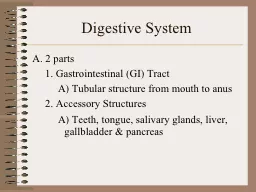
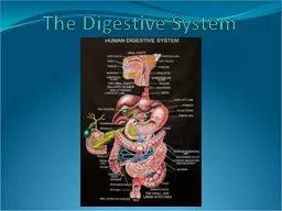
1 Gastrointestinal GI Tract A Tubular structure from mouth to anus 2 Accessory Structures A Teeth tongue salivary glands liver gallbladder amp pancreas Digestive System B 6 basic processes ID: 908738
Download Presentation The PPT/PDF document "Digestive System A. 2 parts" is the property of its rightful owner. Permission is granted to download and print the materials on this web site for personal, non-commercial use only, and to display it on your personal computer provided you do not modify the materials and that you retain all copyright notices contained in the materials. By downloading content from our website, you accept the terms of this agreement.

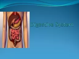
Slide1
Digestive System
A. 2 parts
1. Gastrointestinal (GI) Tract
A)
Tubular
structure from mouth to anus
2. Accessory Structures
A)
Teeth
, tongue, salivary glands, liver, gallbladder & pancreas
Slide2Digestive System
B. 6 basic processes
1.
Ingestion
2.
Secretion
3.
Propulsion
4.
Digestion
(catabolism
)
Slide3Digestive System
A) Mechanical digestion
1
)
Chewing
, mixing with tongue, churning in stomach, segmentation in small intestine
,
and
haustral churning in large intestine
B)
Chemical digestion
1)
Breakdown
by enzymes
5.
Absorption
6.
Defecation
Slide4Digestive System
C. Anatomy of the Digestive System
1. O
ral
cavity (mouth)
A)
Oral
orifice – opens to outside
B)
Lips
& cheeks
1)
Make
up anterior and lateral walls of oral cavity
2)
Composed of a core
of skeletal muscle covered by skin
Slide5Digestive System
3)
Lined
with
nonkeratinized
stratified squamous
4)
Aid
in chewing, keeping food within oral cavity,
and speech
C)
Palate
1)
Forms
superior aspect of the oral cavity (roof of mouth)
2) 2 distinct
portions
a)
Hard
palate – anterior portion
Slide6Digestive System
i
)
Composed
of the maxilla & palatine bones
ii)
Tongue
forces food against it during chewing
b)
Soft
palate – posterior portion
i
)
Soft
, mobile flap that raises to
block the nasopharynx
during swallowing
ii)
Composed
of a core of skeletal muscle
iii)
Uvula
– finger-like projection of the soft palate; function unclear
Slide7Digestive System
D)
Tongue
1)
Makes
up inferior aspect of oral cavity
2)
Moves
food around during mastication (chewing) and swallowing
3)
Essential
for speech production
4)
Contains
taste buds – receptors for various food taste sensations
5)
Lingual
frenulum – connect tongue to floor of mouth
Slide8Digestive System
6)
Papillae
– small elevations on surface of tongue
a)
Aid
in handling of food in mouth
b)
Contain
taste buds & touch receptors
c) 3 types of papillae
i
)
F
iliform
– cone-shaped
(a)
Most
numerous
(b)
Sensitive
to touch
Slide9Digestive System
ii)
Fungiform
– mushroom-shaped
(a) T
aste buds
located on top of the papillae
iii) C
ircumvallate
– resemble fungiform but larger with surrounding furrow
(a) T
aste buds
located on the sides of the papillae
Slide10Digestive System
2.
Salivary glands – produce saliva (pH = 6.75-7.0)
A)
Intrinsic
salivary glands – small
1)
Scattered
within mucosa of tongue, palate, lips & cheeks
2)
Secrete
saliva to keep mouth
moist
Slide11Digestive System
B) Extrinsic salivary glands – large
1) Lie external to oral cavity & secrete saliva into ducts leading to mouth
2) Only secrete saliva as we eat; contains digestive enzymes
Slide12Digestive System
3
) 3 types
a)
Parotid
– lies
slightly anterior & inferior to the ear
b) S
ubmandibular
– on medial surface of mandible, just anterior to mandibular angle
c)
Sublingual
– floor of mouth just inferior to tongue
Slide13Digestive System
3. T
eeth
A) 2 categories
1)
Deciduous
(baby teeth) – 20
a)
Lost
between ages of 6 & 12
2)
Permanent
– 32 (including 3rd molars – wisdom teeth)
Slide14Digestive System
B) 4 types
1) I
ncisors
(8) – chisel-shaped
2)
Canines
(4) – cone-shaped
3)
Premolars
(bicuspids) (8) – broad crown (top) with two rounded cusps (bumps)
4)
Molars
(12) – broad crown & four rounded cusps
Slide15Digestive System
C)
Vascular
& innervated
D)
Tooth
structure
1)
Crown
a)
Portion
above gum (gingiva)
b)
Covered
in enamel
2)
Root
a)
Embedded
in jaw
Slide16Digestive System
b)
Covered
by
cementum
– calcified connective tissue
i
)
Attaches
tooth to the
periodontal ligament
– connects tooth to jaw
3)
Neck
a)
Narrowed
region between crown & root
4)
Dentin
a)
Bone
-like substance; makes up majority of the tooth
Slide17Digestive System
5)
Pulp
cavity
a
)
Located within
the dentin
; houses blood vessels & nerves
6)
Root
canal
a)
Extends
from pulp cavity to proximal end of tooth
; passageway for blood vessels & nerves
7)
Apical
foramen
a)
Opening
at the proximal end of the tooth; allows blood vessels & nerves to enter and leave the tooth
Slide18Slide19Digestive System
4.
Pharynx
A)
Passageway
from mouth to esophagus;
smooth muscles
within propel
food towards the esophagus
B)
Oropharynx
– portion connected to oral cavity
C)
Laryngopharynx
– portion connected to larynx &
esophagus
Slide20Digestive System
5.
E
sophagus
A) Passageway for food from pharynx to stomach
B) Associated structures
1) Esophageal hiatus – passageway through the diaphragm
2) Cardiac orifice – opening between esophagus and stomach
Slide21Digestive System
3
)
Cardiac
sphincter – muscle that closes off to prevent backflow from stomach into
esophagus
C) Lined with
nonkeratinized
stratified squamous
Slide22Digestive System
6.
Stomach
A) Lined with simple columnar epithelium
B)
R
egions
1)
Cardiac
region – encircles the cardiac orifice at junction w/ esophagus
2)
Fundus
– dome-shaped, tucked under
diaphragm
Slide23Digestive System
3) Body – large mid-portion of stomach
4
)
Pyloric
region – terminal region of stomach
a)
Pyloric
sphincter – controls entry of chyme (food) into S.I.
C
)
R
ugae
– longitudinal folds in the mucosa
Slide24Slide25Digestive System
D) Within the wall are a large number of
gastric glands (pits)
1
)
Produce
gastric
juice (pH = 1.5-3.5)
2)
Contain
4 cells types
a)
Goblet
cells – produce an acidic mucus unique to the stomach
b)
Parietal
cells – produce HCl
-
c)
Chief
cells – produce pepsinogen (inactive form of pepsin)
Slide26Digestive System
d)
E
nteroendocrine
cells – produces gastrin
i
)
Released
when food enters the stomach
ii)
Stimulates
the secretion of
HCl
-
and pepsinogen
Slide27Digestive System
7.
Small
intestine
A)
Longest
part of tubular gut (6-7 meters relaxed, 2-4 meters normally)
B) P
ossess
villi
– finger-like projections of the mucosa
1)
Contain
capillaries and
lacteals
Slide28Digestive System
C) Lined with simple columnar epithelium; ciliated with goblet cells
1) Possess
microvilli
– finger-like projection of the columnar cells
D) 3 segments
1) Duodenum (~5%)
a) Receives pancreatic enzymes via the main pancreatic duct, bile via the bile duct, and chyme from the pyloric region of the stomach
Slide29Digestive System
2
)
Jejunum
(~ 40%)
3)
Ileum
(~
55%)
a)
Empties
into the
cecum (large intestine)
Slide30Digestive System
8.
Large
intestine
A) L
ined with simple columnar epithelium; ciliated with goblet cells
B) Subdivisions
1)
Cecum
– sac-like portion inferior to ileocecal valve
a)
Ileocecal
valve – located at junction of ileum and cecum; control movement of
chyme
into L.I.
Slide31Digestive System
2)
Colon
– composed of sac-like pockets known as
haustra
a)
Ascending
colon – moves upward along right posterior abdominal wall up to kidney
b) T
ransverse
colon – extends to the left across abdominal cavity
Slide32Digestive System
c)
Descending
colon – moves downward along left posterior abdominal wall
d)
Sigmoid
colon – S-shaped terminal end of
colon
Slide33Digestive System
3) Rectum – passageway from sigmoid colon to anal canal (anus)
a)
A
nal
canal (anus) – terminal portion of L.I.
i
)
Opens
to outside of body
(a)
Internal
anal sphincter – smooth muscle
(b)
External
anal sphincter – skeletal muscle
Slide34Slide35Digestive System
9.
Liver
A)
Largest
gland in the body
B)
Produces
bile – green, alkaline (basic) liquid stored in the gallbladder
1)
Partially
a digestive product & partially an excretory product
a)
Bile
salts – necessary for lipid digestion & absorption
b)
Bilirubin
– created by the breakdown of RBC
Slide36Digestive System
C) 2 surfaces
1)
Diaphragmatic
(anterior)
– divided into 2 lobes by the
falciform
ligament
a)
Right
lobe
b)
Left
lobe
2)
Visceral
(posterior)
a)
Caudate
lobe – superior
b)
Quadrate
lobe – inferior
Slide37Slide38Digestive System
D) Hepatic artery – carries oxygenated blood from heart to liver
E) Hepatic vein – carries deoxygenated blood from liver to heart
F)
H
epatic
portal vein – carries blood from stomach & intestines to
liver
Slide39Digestive System
G)
Hepatic
ducts – carry bile
1)
Right
– from right lobe
2)
Left
– from left lobe
3)
Common
hepatic – created by a merging of the right & left hepatic ducts
10.
Gallbladder
A)
Small
(~ 4 inches) sac located on the visceral surface of the liver
Slide40Digestive System
B) L
ined with simple columnar epithelium
C) Stores & concentrates bile
D) Cystic
duct – carries bile to & from gallbladder
1)
Merges
with common hepatic duct to form the bile duct
2)
Bile
duct – carries bile to duodenum
Slide41Slide42Digestive System
11.
Pancreas
A)
Endocrine
& exocrine organ
B)
Tadpole
-shaped
1)
Consists
of a head, body & tail
C)
Produces pancreatic juice (pH = 8.0)
1) Contains many
of the enzymes used by the
S.I.
for
digestion
Slide43Digestive System
2) Produced by the aciner cells
a)
A
cini
– clusters of aciner cells
D)
A
ccessory
pancreatic duct – lies at head on pancreas and merges with main pancreatic duct
E
)
M
ain
pancreatic duct – merges with bile duct and empties into the
duodenum
1
) Release of bile & pancreatic juice controlled by
hepatopancreatic
sphincter
Slide44Slide45Digestive System
D. Digestion & Absorption
1. M
outh
A)
Mechanical
digestion
1)
Chewing
and rolling of food into bolus
B)
Chemical
digestion
1)
Salivary
amylase
a)
Starts
breakdown of starch
Slide46Digestive System
2)
Lingual
lipase
a)
Starts
breakdown of dietary triglycerides
C)
Normally
no
absorption occurs
D)
Swallowing
1) 3 phases
a)
Voluntary
phase
b)
Pharyngeal
phase
c)
Esophageal
phase
Slide47Digestive System
2. Esophagus
A)
Enzymes
from the mouth are still working
B)
Secretes
no enzymes only mucus
C)
No
absorption
D)
Peristalsis
1)
Wave-like
, smooth muscle contractions that move foodstuffs through the GI tract
Slide48Digestive System
3. Stomach
A)
Mechanical
digestion
1)
Churning
(smooth muscle contractions)
mixes bolus with gastric juices yielding
chyme
Slide49Digestive System
B) Chemical digestion
1) HCl
-
a
)
Inactivates
salivary amylase & lingual lipase
b)
Initiates
protein catabolism by unfolding protein structure & activating
pepsin
Slide50Digestive System
2) Pepsin
a) Produced when HCl
-
activates pepsinogen
b) Begins breakdown of peptide bonds
Slide51Digestive System
C)
Very
little absorption
1) W
ater
, ions, aspirin,
and
alcohol
D)
Releases
chyme
into
S.I.
in small amounts over a period of time (~4 hours)
4. Small Intestine
A)
Mechanical
digestion
1)
Peristalsis
Slide52Digestive System
2)
Segmentation
a)
Oscillating
, ring-like, smooth muscle contractions
i
)
Mixes
chyme with digestive juices
ii)
Brings
digestive products into contact with mucosa helping absorption
B)
Chemical
digestion
1) CHO catabolism – desirable end product is glucose in all cases (however, sometimes the end product is fructose or
galactose
)
Slide53Digestive System
a)
Brush
border
enzymes (released from the small intestine itself)
i
)
G
lucoamylase
ii)
D
extrinase
iii)
Maltase
iv)
S
ucrase
v)
Lactase
b)
Pancreatic
enzymes
i
)
Pancreatic
amylase
Slide54Digestive System
2)
Protein
catabolism – desirable end product is a single amino acid
a)
Brush
border enzymes
i
)
Carboxypeptidase
ii)
A
minopeptidase
iii)
D
ipeptidase
Slide55Digestive System
b) Pancreatic enzymes
i
)
Trypsin
& chymotrypsin
ii)
C
arboxypeptidase
Slide56Digestive System
3) Lipid catabolism – desirable end products are 2 fatty acids & 1
monoglyceride
or 3 fatty acids and 1 glycerol
a) Bile salts
i
) Emulsification
b) Pancreatic lipase
Slide57Digestive System
B)
Absorption
– about 90% of all absorption occurs here
1) CHO
absorption (monosaccharides; glucose, fructose, galactose)
a)
Fructose
i
)
Facilitated
diffusion
b)
Glucose
& galactose
i
)
Secondary
active transport with Na
+
(
cotransport
)
Slide58Digestive System
2) Protein absorption (individual amino acids)
a) Secondary active transport with Na
+
(
cotransport
)
Slide59Digestive System
3
)
Lipid absorption (monoglycerides & fatty acids)
a)
Aided
by the actions of bile
i
)
B
ile
salts
& lecithin bind with fatty acids & monoglycerides forming small clusters known as
micelles
(a)
Micelles
are absorbed into the columnar cells where triglycerides reform
Slide60Digestive System
ii)
Triglycerides
are coated with phospholipids & cholesterol resulting in
chylomicrons
iii)
Chylomicrons
are then absorbed into the
lacteals by simple diffusion
C)
Chyme may
spend up to 4 hours in the small intestine
Slide61Digestive System
5. Pancreas
A)
Accessory
to
the S.I.
B)
Produces
:
1)
Pancreatic
juice
a)
Buffers
the acidic chyme coming from the stomach
i
)
Stops the action
of pepsin
2) P
ancreatic
enzymes
a)
Work
in small intestine
Slide62Digestive System
6. Liver
A)
Accessory
to
the S.I.
B)
Many
functions
1)
Plasma
protein production
2)
Removal
of drugs & hormones
3)
Fat
-soluble vitamin storage
4)
Produces & stores
glycogen
Slide63Digestive System
5)
Phagocytosis
of
worn/old RBC
a)
Results
in the production of bilirubin
6)
Synthesis
of bile salts
7)
Produces
bile
a)
Yellow
-green, alkaline solution containing bile salts, bilirubin, cholesterol, lecithin & and a number of electrolytes
b)
Involved
with lipid catabolism & absorption
Slide64Digestive System
7. Gallbladder
A)
Stores
& concentrates bile by absorbing water & ions
B)
Releases
bile into
S.I.
in response to the release of
cholecystokinin (CCK)
1)
Released
from intestinal lining in response to fatty chyme entering the
duodenum
Slide65Digestive System
8. Large Intestine
A
)
Digestion
1)
Mechanical digestion
a)
Peristalsis
at a slow rate
b)
Haustral
churning
i
)
Contraction
of an individual
haustrum
Slide66Digestive System
c) Mass peristalsis
i
) Strong wave beginning in transverse colon and pushing contents into rectum
Slide67Digestive System
2)
Chemical digestion
a)
No enzymes secreted, just mucus
b)
Bacteria
living in
L.I.
finish digestion
i
)
Ferment
CHO
–
provide
themselves with energy
ii)
Some
B vitamins & vitamin K are end products of bacterial action
Slide68Digestive System
B)
Absorption
1)
Water
2)
Electrolytes
(Na
+
&
Cl
-
)
3)
Vitamins
C) C
hyme
may remain in the large intestine for 3-10 hours
Slide69Digestive System
D) Defecation
1)
Lumbar
reflex initiated when feces enters the rectum
2)
Impulses
travel back to internal anal sphincter as well as to the cerebral cortex
a)
Internal
anal sphincter relaxes allowing feces into the anus
3)
Cerebral
cortex fires causing external anal sphincter to
relax and the rectal muscles to contract
Slide70Digestive System
E. Disorders of the Digestive System
1. Peritonitis – inflammation of the peritoneum
2. Mumps – swollen parotid glands as a result of a virus (Myxovirus)
3. Heartburn – failure of the cardiac sphincter to remain closed
4. Hiatal hernia – upper portion of the stomach protrudes above the diaphragm
Slide71Digestive System
5. Gastric (or Peptic) ulcers – erosion of the stomach (or small intestine) wall associated with the
Helicobacter
bacteria
6. Enteritis – inflammation of either intestine; however usually the small intestine
7. Hepatitis – inflammation of the liver as a result of a viral infection (A-E, G)
Slide72Digestive System
8. Cirrhosis – chronic inflammation of the liver due to alcoholism or hepatitis
9. Gallstones – highly concentrated cholesterol derivatives in bile
10. Jaundice – accumulation of bilirubin in the skin as a result of a blockage or liver disease resulting in a yellow skin color