
and their associated extracellular substancesECM which are specialized to carry out specific functions B Four Primary Tissues of the Body 1 Epithelium 2 Connective Tissue 3 Muscle ID: 920572
Download Presentation The PPT/PDF document "TISSUE A. Definition: societies of cel..." is the property of its rightful owner. Permission is granted to download and print the materials on this web site for personal, non-commercial use only, and to display it on your personal computer provided you do not modify the materials and that you retain all copyright notices contained in the materials. By downloading content from our website, you accept the terms of this agreement.


Slide1
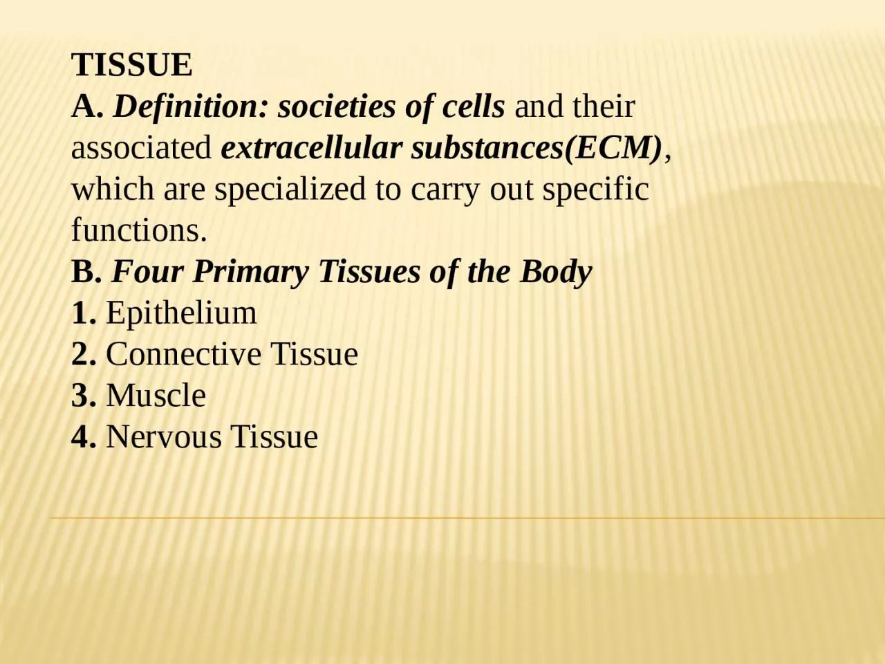
TISSUE
A.
Definition: societies of cells
and their associated
extracellular substances(ECM)
, which are specialized to carry out specific functions.
B.
Four Primary Tissues of the Body
1.
Epithelium
2.
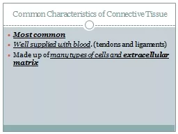
Connective Tissue
3.
Muscle
4.
Nervous Tissue
Slide2Slide3Slide4Epithelium is one of the four basic types of animal tissue, along with connective tissue,
muscle tissue
and
nervous tissue
. Epithelial tissues line the
cavities
and surfaces of structures throughout the body. Many glands are made up of epithelial cells. Functions of epithelial cells include secretion, selective absorption, protection, Epithelial layers contain no blood vessels, so they must receive nourishment via diffusion of substances from the underlying connective tissue, through the
basement membrane
.
Slide5In general, tissues are classified by the morphology of their cells, and the number of layers they are composed of. Epithelial tissue that is only one cell thick is known as simple epithelium. If it is two or more cells thick, it is known as stratified epithelium. However, when taller simple epithelial cells (see
columnar
, below) are viewed in cross section with several nuclei appearing at different heights, they can be confused with stratified epithelia. This kind of epithelium is therefore described as
"
pseudostratified
"
epithelium.
Classification of epithelia
1.
Surface epithelia
2.
Glandular epithelia
3.
Special epithelia
Slide6Functions of EpitheliaMajor functions of epithelia include:1. Protecting underlying structures. Examples include the skin and the epithelium of the oral cavity, which protects the underlying structures from abrasion.2.
Acting as barriers.
Epithelium prevents the movement of many substances through the epithelial layer. For example, the skin acts as a barrier to water and prevents water loss from the body.
Slide73. Permitting the passage of substances. Epithelium allows the movement of many substances through the epithelial layer. For example, oxygen and carbon dioxide are exchanged between the air and blood by diffusion through the epithelium in the lungs.4.
Secreting substances.
Examples include the sweat glands, mucous glands, and the enzyme-secreting portion of the pancreas.
5.
Absorbing substances.
The cell membranes of certain epithelial tissues contain carrier molecules that regulate the absorption of materials.
Slide8Cell ConnectionsLateral and basilar surfaces have structures that serve to hold cells to one another or to the basement membrane .These structures do three things: (1) they mechanically bind the cells together,(2) they help form a permeability barrier, and (3) they providea mechanism for intercellular communication. Epithelial cells secrete
glycoproteins
that attach the cells to the basement membrane and to one another. This relatively weak binding between cells is reinforced by
desmosomes
,disk-shaped structures with especially adhesive
glycoproteins
that bind cells to one another and intermediate filaments that extend into the cytoplasm of the cells. Many
desmosomes
are found in epithelia that are subjected to stress, such as the stratified
squamous
epithelium of the skin.
Hemidesmosomes
,
similar to one-half of a
desmosome,attach
epithelial cells to the basement membrane
Slide9Important characteristics1. Covers or lines all body surfaces2.
Polarity (epithelial cells have specific apical, lateral & basal domains)
3.
Characterized by specific intermediate filaments called
cytokeratins
4.
Attached to underlying CT via ECM basement membrane
5.
Generally
avascular
6.
Gives rise to majority of glands
7.
High regenerative capacity; plasticity
8.
Great diversity of function
Slide10Simple epitheliumType
Squamous
Squamous
cells have the appearance of thin, flat plates. They fit closely together in tissues; providing a smooth, low-friction surface over which fluids can move easily. The shape of the nucleus usually corresponds to the cell form and helps to identify the type of epithelium.
Squamous
cells tend to have horizontally flattened, elliptical (oval or shaped like an egg) nuclei because of the thin flattened form of the cell. Classically,
squamous
epithelia are found lining surfaces utilizing simple passive diffusion such as the
alveolar epithelium
in the lungs. Specialized
squamous
epithelia also form the lining of cavities such as the blood vessels (
endothelium
) and
pericardium
(
mesothelium
) and the major cavities found within the body.
Slide11Cuboidal As their name implies, cuboidal cells are roughly cuboidal
in shape, appearing square in cross section. Each cell has a spherical nucleus in the centre.
Cuboidal
epithelium is commonly found in secretive or absorptive tissue: for example the (secretive) exocrine gland the pancreas and the (absorptive) lining of the kidney tubules as well as in the ducts of the glands. They also constitute the
germinal epithelium
that covers the female ovary.
Slide12Columnar Columnar epithelial cells are elongated and column-shaped. Their nuclei are elongated and are usually located near the base of the cells. Columnar epithelium forms the lining of the stomach and intestines. Some columnar cells are specialized for sensory reception such as in the nose, ears and the taste buds of the tongue. Goblet cells (unicellular glands) are found between the columnar epithelial cells of the duodenum. They secrete mucus, which acts as a lubricant.
Slide13Pseudostratified These are simple columnar epithelial cells whose nuclei appear at different heights, giving the misleading (hence "pseudo") impression that the epithelium is stratified when the cells are viewed in cross section. Pseudostratified epithelium can also possess fine hair-like extensions of their apical (luminal) membrane called
cilia
. In this case, the epithelium is described as "ciliated"
pseudostratified
epithelium. Cilia are capable of energy dependent
pulsatile
beating in a certain direction through interaction of
cytoskeletal
microtubules and connecting structural proteins and enzymes. The wafting effect produced causes mucus secreted locally by the goblet cells (to lubricate and to trap pathogens and particles) to flow in that direction (typically out of the body). Ciliated epithelium is found in the airways (nose, bronchi), but is also found in the uterus and Fallopian tubes of females, where the cilia propel the ovum to the uterus.
Slide14Slide15Slide16Stratified epitheliumStratified epithelium differs from simple epithelium in that it is multilayered. It is therefore found where body linings have to withstand mechanical or chemical insult such that layers can be abraded and lost without exposing subepithelial layers. Cells flatten as the layers become more apical, though in their most basal layers the cells can be squamous
,
cuboidal
or columnar.
Stratified epithelial tissue also differs from simple epithelial tissue in that stratified epithelial tissues do not contain
junctional
complexes, and have their cells bound together only by
desmosomes.Stratified
epithelia (of columnar,
cuboidal
or
squamous
type) can have the following specializations
Slide17SpecializationDescriptionKeratinizedIn this particular case, the most apical layers (exterior) of cells are dead and lose their nucleus and cytoplasm, instead contain a tough, resistant protein called keratin. This specialization makes the epithelium waterproof, so is found in the mammalian skin. The lining of the esophagus is an example of a non-keratinized or "moist" stratified epithelium.
Transitional
Transitional epithelia are found in tissues that stretch and it can appear to be stratified
cuboidal
when the tissue is not stretched or stratified
squamous
when the organ is distended and the tissue stretches. It is sometimes called the
urothelium
since it is almost exclusively found in the bladder,
ureters
and urethra.
Slide18GLANDULAR EPITHELIA Glands are formed from the invagination/infolding
of epithelium and subsequent growth in the underlying connective tissue. There are two major classifications of glands:
endocrine glands
and
exocrine glands
. Endocrine glands secrete their product into the extracellular space where it is rapidly taken up by the blood vascular system. Exocrine glands secrete their products into a duct that then delivers the product to the lumen of an organ or onto the free surface of the epithelium.
A.
Classification
1.
Types of glands
a.
Exocrine
b.
Endocrine (
hemocrine
)
c.
Paracrine
d.
Autocrine
Slide19Slide20B.Ducts: structure in exocrine glands1. Simple glands (=unbranched ducts)2. Compound glands (=branched ducts)
C.
Secretory
components
1.
Number of
secretory
cells
a.
Unicellular (e.g., goblet cells)
b.
Multicellular
glands (majority)
2.
Structure of
secretory
units
a.
Tubular
b.
Acinar
/alveolar
Slide21D. Types of secretion (secretory product)1. Mucous2. Serous
3.
Seromucous
E.
Mechanisms of secretion
(methods of release)
1.
Merocrine
(e.g., Salivary
glands,Pancreas
)
2.
Apocrine
(e.g.,
Apocrine
sweat glands,
Mammary glands)
3.
Holocrine
(e.g., Sebaceous glands )
Slide22Myoepithelial cells1. Contractile, branched epithelial cells2. Located between glandular epithelial cells and basement membrane3.
Contain
actin
filaments; smooth muscle-like
4.
Facilitate glandular secretion.
Slide23