
left lower quadrant mass causes 1Skin Sebaceous cyst malformation Abscess inflammation Primary and metastatic carcinomas neoplasm Contusiontruma 2Subcutaneous Tissue and Fascia Hernia ID: 1007569
Download Presentation The PPT/PDF document "Left lower quadrant abdominal pain and m..." is the property of its rightful owner. Permission is granted to download and print the materials on this web site for personal, non-commercial use only, and to display it on your personal computer provided you do not modify the materials and that you retain all copyright notices contained in the materials. By downloading content from our website, you accept the terms of this agreement.


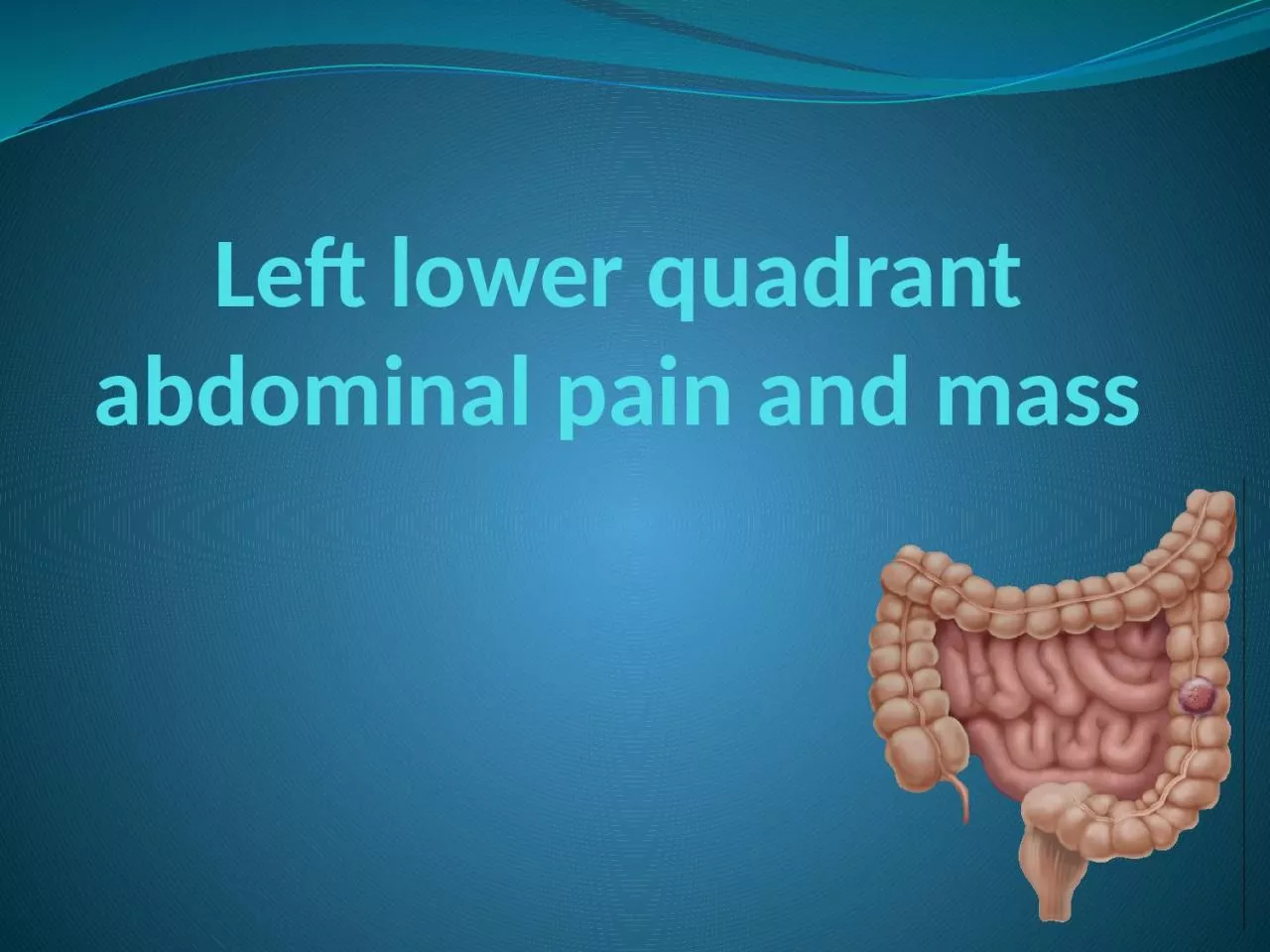
1. Left lower quadrant abdominal pain and mass
2. left lower quadrant mass causes1-SkinSebaceous cyst (malformation)Abscess (inflammation)Primary and metastatic carcinomas (neoplasm)Contusion(truma)2-Subcutaneous Tissue and FasciaHerniaCellulitisMetastatic carcinoma ⇧ Contusion Lipoma3-Muscle Myositis Contusion
3. left lower quadrant mass causes4-Sigmoid ColonDiverticulumDiverticulitis and abscessCarcinoma and polypPerforation Volvulus Contusion Intestinal obstructionTuberculosis Foreign body Granulomatous and ulcerative colitis
4. left lower quadrant mass causes5-Tube and OvaryHydatid cyst of MorgagniTubo-ovarian abscessOvarian cyst and carcinoma Ectopic pregnancy 6-Iliac Artery and Veins and AortaAneurysmThrombophlebitis
5. left lower quadrant mass causes7-Lymph Nodes Tuberculous and acute infectious adenitisMetastatic tumor8-IliumOsteomyelitisSarcomaFracture or contusion
6. Left lower quadrant pain causes 1-Abdominal abscess2. Colonic volvulus3. Constipation4. Crohn's disease5. Cystitis6. Diverticular Disease7. Ectopic pregnancy8. Inguinal hernia9. Intussusception10. Ovarian cysts11. Pelvic Inflammatory Disease12. Pelvic abscess13. Rectal abscess14. Tuberculosis15. Ulcerative colitis16. Ulcerative proctosigmoiditis17. Uterine fibroids
7. Colorectal cancer* Includes cancerous growths in the colon, rectum and appendix.*Precursor of ~90% of colorectal cancers is the adenomatous polyp. These mushroom-shaped growths are usually benign, but some develop into cancer over time. *Very curable if detected in early stage
8. adenomatous polypcolorectal cancers
9. Colorectal cancer*The transition from normal mucosa to polyp to invasive cancer is usually a lengthy process (7 – 12 years in many cases). Polyp size correlates to cancer probabilityPolyps < 1 cm 1% are cancerousPolyps > 2 cm 30% are cancerous
10. Human colon carcinogenesis progresses by the dysplasia/adenoma to carcinoma pathway
11. Epidemiology*There are nearly one million new cases of colorectal cancer diagnosed world-wide each year and half a million deaths.*Most frequent form of cancer among persons aged 75 years and older.*It is the fifth most common form of cancer in the United States and the third leading cause of cancer-related death in the Western world.
12. Epidemiology 30% - 50% of population will develop adenomatous polyps over lifetime1% - 3% of polyps become malignantMost remain asymptomatic & undetectedPrevalence of polyps increases with age50% of men, 40% of women by age 50> 90% of CRC diagnosed after 55 yrs
13. pathologyColorectal cancer is a disease originating from the epithelial cells lining the colon or rectum , as a result of mutations along the 'Wnt signaling pathway. Some of the mutations are inherited, and others are acquired. The most commonly mutated gene in all colorectal cancer is the APC gene, which produces the APC protein.
14. The malignant potential of a polyp based on histology is: tubular < tubulovillous < villous. Cancer risk also rises with increasing size of the polyp. The table below shows the incidence of invasive carcinoma related to polyp histology and size based on analysis of 7000 polypectomy specimens. Colon Cancer ModuleBackTubularTubulovillousVillous size cm0.5-0.9 0.3% 1.5% 2.5%1.0-1.9 3.6% 6.4% 5.7%2.0-2.9 6.5% 11.4% 17.0%>3.0 11.0% 16.0% 20.0%Colonic polyps article Images from: www.endoatlas.com/atlas_co.html
15. Colon Cancer ModuleBackHyperplastic PolypsThe epithelial cells at the base of the crypt (regenerative zone) have mildly enlarged, but uniform nuclei and brisk mitotic rate, feature which is normally present in reactive colonic mucosa Endoscopic image of hyperplastic polypswww.GI-Pathology.netwww.GI-Pathology.net
16. Colon Cancer ModuleTubular AdenomaBack The adenomatous polyp has a smooth outline and is composed of numerous architecturally simple crypts with mild irregularity in size and shape Endoscopic image of a tubular adenoma www.GI-Pathology.netwww.endoatlas.com/atlas_co.html
17. Colon Cancer ModuleVillous AdenomaBackVillous adenoma with glands that extend straight down from the surface to the base as fingerlike projectionsEndoscopic image of a villous adenomawww.endoatlas.com/atlas_co.htmlwww.GI-Pathology.net
18. Colon Cancer ModuleTubulovillous AdenomaBackTubulovillous adenoma with 80% tubular histology and 20% villous histologyEndoscopic image of a tubulovillous adenoma www.endoatlas.com/atlas_co.htmlwww.GI-Pathology.net
19. The most common colon cancer cell type is adenocarcinoma which accounts for 95% of cases. Other, rarer types include lymphomaand squamous cell carcinoma.
20. Local symptoms*change in bowel habit*feeling of incomplete defecation (tenesmus)and reduction in diameter of stoolrectal cancer.*passage of bright red blood in the stool. *mucus*Melena when the disease is located in the beginning of the large bowel.
21. Local symptomsLarge tumor:*Bowel obstruction(constipation, abdominal pain, abdominal distension, vomiting, perforation and peritonitis)*Noticed by physical examination.
22. Local symptomsLocal invasion:-blood or air in the urine (invasion of the bladder)-vaginal discharge (invasion of the femalereproductive tract).
23. Constitutional symptoms *chronic occult bleeding cause iron deficiency anemia fatigue, palpitations and pallor. *decreased appetite and wieght loss. paraneoplastic syndrome:*Fever(unusual)*thrombosis, usually deep vein thrombosis (most common)
24. Metastatic symptomColorectal cancer most commonly spreads to the liver jaundice , abdominal pain.
25. Risk factorAverage risk”AGE: population > 50 years~ 75% of CRC cases occur in this group *Before 50 uncommon unless a family history of early colon cancer.
26. Risk factorHigh risk”Factors include personal and/or familial history of CRC or polyps, genetic syndromes (hereditary CRC), history of inflammatory bowel disease ~ 25% of CRC cases occur in this group
27. Risk factorHereditary: Familial adenomatous polyposis (FAP) carries a near 100% risk of developing colorectal cancer by the age of 40 if untreatedHereditary nonpolyposis colorectal cancer (HNPCC) or Lynch syndrome
28. Risk factorInflammatory bowel disease: About one percent of colorectal cancer patients have a history of chronic ulcerative colitis. Crohn's disease have a risk of colorectal cancer, but less than that of patients with ulcerative colitis.
29. Colon Cancer ModuleInflammatory Bowel Disease Ulcerative Colitis & Crohn’s Colitiswww.GI-Pathology.netwww.GI-Pathology.netUlcerative Colitis gross specimenCrohn’s Colitis endoscopic view Barium enema: colon with "lead-pipe" appearance in ulcerative colitis www.learningradiology.comBack
30. Familial Adenomatous Polyposis
31. Risk for Colorectal CancerDietary factors*High fat diets correlate w/ high rates of CRC*Protective effects of fiber are still unproven*Excessive calorie intake may enhance riskBehavioral factors*Physical activity may reduce risk*Excessive alcohol use and smoking have been associated with increased risk
32. Colorectal cancer risk*Women who have had cancer of the ovary, uterus, or breast are at higher risk of developing colorectal cancer.*Exogenous estrogens such as hormone replacement therapy (HRT), tamoxifen, or oral contraceptives might be associated with colorectal tumors *Lifestyle changes could decrease the risk ofcolorectal cancer as much as 60-80%.
33. Early Detection of Colorectal CancerDetecting and removing polyps has been shown to reduce incidence of CRCCommonly used screening tests includeDigital Rectal ExamDouble-Contrast Barium Enema (DCBE)Fecal Occult Blood Test (FOBT)Flexible Sigmoidoscopy (FS)Colonoscopy
34. Digital Rectal Examination (DRE)*Not effective as CRC screening test*Sensitivity for CRC less than 10%*Often used to obtain stool sample for FOBT with chance for increased false positive results
35. Double-Contrast Barium Enema*Radiologic study—barium is used as contrast material to visualize lumen of the colon*Effectiveness as screening test debated*National Polyp Study: Sensitivity of 48% for polyps > 1 cm*Further evaluation required if polyps detected*Less risk of perforation than endoscopic exams*May be recommended with flexible sigmoidoscopy for CRC screening
36.
37. This is an x-ray called barium enema. The area in the circle shows an advanced cancer of colon that has produced an apple core lesion. The name apple core comes from the x-ray appearance of circumferentially eaten apple.
38. This represents the infiltration of the cancer into the muscular wall of the colon and the consequent loss of elasticity of the colon itself
39. Double-Contrast Barium EnemaRectal cancerColon cancer
40. Fecal Occult Blood Testing (FOBT)*Detects blood from cancers or large polyps*Bleeding is intermittent and increases with polyp size and stage of cancer*Hemoccult II (guaiac-based) most widely used (0ne of FOBT type)*Inexpensive and easy to perform *Diet and medications affect resultsFalse positives: oral iron, aspirin, NSAIDs, anticoagualantsFalse negatives: vitamin C*33% CRC mortality reduction with annual screening
41. SigmoidoscopyAdvantagesRelatively accurate Quick procedure performed w/out sedationInexpensive
42.
43. SigmoidoscopyDisadvantages *Misses 40% - 50% of CRC and polyps *Risk of colon perforation is 1 to 2 per 10,000 exa*Evidence for most effective screening interval is inconclusive
44. Colonoscopy*95% of CRC in reach of colonoscope*could eliminate 80% to 90% of CRC mortality in population over age 50 years*Diagnostic use after positive results on FOBT or FS *Recommended as initial screening test for high risk individuals
45. Polyps can be detected and removed during colonoscopy. Studies show this procedure would decrease by > 80% the risk of cancer death, started by the age of 50, and repeated every 5 or 10 years.
46. ColonoscopyDisadvantagesExpensiveNegative impact on patient’s daily lifeTrained endoscopists must performBowel perforation most serious complication1 to 3 per 1000 procedures
47. CRC Screening Guidelines Screening should be individualized according to age and comorbiditiesThree screening options suggested: *Double-contrast barium enema (DCBE) plus FS every 5 years*Colonoscopy every 10 years*FOBT annually and flexible sigmoidoscopy (FS) every 5 year.
48. MonitoringCarcinoembryonic antigen (CEA) is a protein found on virtually all colorectal tumors. CEA may be used to monitor and assess response to treatment in patients with metastatic disease. CEA can also be used to monitor recurrence in patients post-operatively.
49. stagingTNM (for tumors/nodes/metastases) system"T" denotes the degree of invasion of the intestinal wall, "N" the degree of lymphatic node involvement, and "M" the degree of metastasis.
50. stagingDukes systemA - Tumour confined to the intestinal wallB - Tumour invading through the intestinal wallC - With lymph node(s) involvement (this is further subdivided into C1 lymph node involvement where the apical node is not involved and C2 where the apical lymph node is involved)
51. TNM stage criteria for colorectal cancer[39]TNM stageAJCC stageTis: Tumor confined to mucosa; cancer-in-situTis N0 M0Stage 0T1: Tumor invades submucosaT1 N0 M0Stage IT2: Tumor invades muscularis propriaT2 N0 M0Stage IT3: Tumor invades subserosa or beyond (without other organs involved)T3 N0 M0Stage II-AT4: Tumor invades adjacent organs or perforates the visceral peritoneumT4 N0 M0Stage II-BN1: Metastasis to 1 to 3 regional lymph nodes. T1 or T2.T1-2 N1 M0Stage III-AN1: Metastasis to 1 to 3 regional lymph nodes. T3 or T4.T3-4 N1 M0Stage III-BN2: Metastasis to 4 or more regional lymph nodes. Any T.any T, N2 M0Stage III-CM1: Distant metastases present. Any T, any N.any T, any N, M1Stage IV
52. TreatmentSurgery remains the primary treatment while chemotherapy and/or radiotherapy may be recommended depending on the individual patient's staging and other medical factors.
53. surgeryCurative palliative bypass fecal diversion open-and-close.
54. polypectomyCurative *tumor is localized.*Very early cancer polypectomy at the time of colonoscopy.
55. curative surgerymore advanced tumor colectomy:removal of the section of colon containing the tumor with sufficient marginsand radical resection of mesentery and lymph nodes to reduce local recurrence. *The remaining parts of colon are anastomosed*If anastomosis not possible, a stoma is created
56. Colon Cancer ModuleLeft HemicolectomyBack
57. Colon Cancer ModuleAbdominoperineal ResectionBack
58. colectomy specimen containing two adenomatou polyps
59. palliative (non curative) resectionIn case of multiple metastasesto reduce further morbidity bleeding, invasion, and catabolic effect *Surgical removal of isolated liver metastases ( common ) with chemotherapy may be curative in selected patients.
60. palliative (non curative) resectionIf the tumor invaded into adjacent vital structures which makes excision technically difficult, the surgeons may prefer to bypass the tumor (ileotransverse bypass) or to do a proximal fecal diversion through a stoma.
61.
62. open-and-closeThe worst case would be an open-and-close surgery, when surgeons find the tumor unresectable and the small bowel involved; any more procedures would do more harm than good to the patient. This is uncommon with the advent of laparoscopy and better radiological imaging. Most of these cases formerly subjected to "open and close" procedures are now diagnosed in advance and surgery avoided.
63. Chemotherapy• Stage III patients benefit most from chemotherapyafter surgery.• Some Stage II patients may also benefit.• Usually given for 6 months.• Old standard was 5-FU/Leucovorin given weekly,or for 5 days in a row once a month.• Oral chemotherapy drug, Xeloda, is equivalent tothe IV 5-FU.• Newly approved drug, Oxaliplatin, given with 5-FU adds additional benefit.
64. Follow-up after Surgery/Chemo• Clinical Exam every 3 months for 2 years,every 6 months for 5 years• Colonoscopy within 1 year of resection• Tumor Markers- (CEA and CA 19-9)substances produced by cancer cells whichare detectable in the blood, may be anyearly indication of recurrent disease• CT scans can be considered for high risk
65. Rectal Cancer• Radiation has a major role• Given either before surgery, or after surgery• Chemotherapy with 5-FU is given by continuousinfusion through an intravenous (IV) line at thesame time as radiation to make the radiation moreeffective.• Studies show that radiation along with surgerydecreases the risk of recurrenceRadiotherapy is not used routinely in colon cancer
66. ProgonsisSTAGE TNM GROUP GROUP DUKE’S Prognosis Stage I T1 N0 M0 Duke’s A 5 year survival >90% T2 N0 M0 Stage II T3 N0 M0 Duke’s B 5 year survival 70-85% T4 N0 M0 5 year survival 55-65% Stage III any T N1 M0 Duke’s C 5 year survival 45-55% any T N2, N3 M0 5 year survival 20-30% Stage IV any T any N M1 (distant) Duke’s D 5 year survival < 5%