
UNIVERSITY OF WARMIA AND MAZURY OLSZTYN PATHOMORPHOLOGY III INDEX INTRODUCTION SYMPTOMS MACROSCOPIC LESIONS MICROSCOPIC LESIONS TREATMENT PREVENTION BIBLIOGRAPHY INTRODUCTION Caused ID: 1037918
Download Presentation The PPT/PDF document "FELINE INFECTIOUS PANLEUKOPENIA" is the property of its rightful owner. Permission is granted to download and print the materials on this web site for personal, non-commercial use only, and to display it on your personal computer provided you do not modify the materials and that you retain all copyright notices contained in the materials. By downloading content from our website, you accept the terms of this agreement.


1. FELINE INFECTIOUS PANLEUKOPENIA UNIVERSITY OF WARMIA AND MAZURY, OLSZTYNPATHOMORPHOLOGY III
2. INDEX INTRODUCTION SYMPTOMS MACROSCOPIC LESIONS MICROSCOPIC LESIONS TREATMENT PREVENTIONBIBLIOGRAPHY
3. INTRODUCTIONCaused by feline parvovirus. Family Parvoviridae, gender Parvovirus.Highly contagious viral diseaseKittens are most severly affected It produces leukopenia = immunosuppressed catsIt is relatively frequent todayIt is subject to vaccination“The feline parvovirus infects and kills cells that are rapidly growing and dividing, such as those in the bone marrow, intestines, and the developing fetus”
4. Ways of infection: Direct contact with urine, stool and nasal secretions Infection by fleas as vectors Transplacental Infected cats shed the virus for a relatively short period of time (1-2 days)The virus can survive for up to a year in the environment = infection without ever coming into direct contact with an infected cat. It is very important to isolate infected cats. Any materials used on or for infected cats should not be used or allowed to come in contact with other cats, and people handling infected cats should practice proper hygiene to prevent spreading the infection.The virus that causes FP is difficult to destroy and resistant to many disinfectants.
5. SYMPTOMS ENTERIC AND ACUTE FORM (KITTENS 2-4 months): Vomiting and diarrhea = dehydration Immunosupression Injuries in small intestine Posible subclinic infectionREPRODUCTIVE FORM:Pregnant females with infection in the uterus Animals that have been infected in the early neonatal period
6. MACROSCOPIC LESIONSExternal evidence of diarrheaThe eyes may be sunkenThe skin is usually inelastic, reflecting dehydration. Rehydrated animals may have edema, hydrothorax, and ascites resulting from hypoproteinemia. Pallor of mucous membranes and internal tissues in anemic animals. Gross lesions of internal organs most consistently involve the thymus and the intestine. The thymus is markedly involute and reduced in mass in young kittens. Enteric lesions may be subtle and easily overlooked.
7. Intestinal serosa may appear dry and nonreflective, with an opaque ground-glass appearance. Petechiae or more extensive hemorrhage in the subserosa, muscularis, or submucosa of the intestinal wall. The content is usually foul-smelling, scant, and watery, and yellow-gray at all levels of the intestine. Lesions over Peyer's patches in the ileum. Formed feces are not evident in the colon. Lymph nodes may be prominent at the root of the mesentery. Gross lesions elsewhere in the carcass are usually restricted to pulmonary congestion and edema in some animals.
8. Dilated and hemorrhagic small intestine in feline panleukopenia.(Couresy J. Cawell)Hemorragic enteritis as a consecuence of feline panleukopenia virus infection(Vet.Pathol.Utrecht)Intestinal damage as a consequence of feline panleukopenia virus infection; sloughing of gut epithelium and fibrinous “casts” are prominent.Vet.Pathol.Utrecht
9. Cerebellar hypoplasia in a kitten infected in utero with feline panleukopenia virus.Marian C. HorzinekCerebellar hypoplasia (below) in a kitten infected in utero with feline panleukopenia virus; a normal brain is shown for comparison (above)Diane Addie
10. A kitten with severe signs of dehydration, a result of electrolyte loss as a consequence of feline panleukopenia.Diane Addie
11. MICROSCOPIC LESIONSLesions in the intestinal tract vary with the severity and duration of the disease.Usual in lymphoid organs and bone marrowCrypt-lining epithelium is infectedIntranuclear inclusions may be foundDamaged epithelium containing inclusions exfoliates into the lumen of crypts. Crypts are dilatedThe lamina propia between crypts contains numerous neutrophils and eosinophilsThe mucosa becomes thin and eroded or ulcerated, with effusion of tissue fluids, fibrin, and erythrocytesInflammatory cells and superficial masses of bacteriaFocal mucosal collapse and erosion or ulceration
12. Colonic lesions are present in about half of fatal cases of panleukopeniaLesions of lymphoid organs during the early phase of the disease consist of lymphocytolysis in follicles and paracortical tissue in lymph nodesLymphoid necrosis has been associated with induced apoptosis of virus-infected lymphocytesFollicular hyalinosis, the presence of amorphous eosinophilic material in the center of depleted follicles, may be seenSeverely depleted Peyer's patches may be difficult to recognize microscopicallyLater in the course of clinical disease, corresponding to the period beyond 7-8 days after infection, prominent regenerative lymphoid hyperplasia may be found.In severely affected animals with leukopenia proliferating elements in the bone marrow may be depleted.
13. Necrosis and dilation of crypts of Lieberkühn, caused by feline panleukopenia virus infection in a catHistological section of feline small intestine diagnosed with feline panleucopenia. Acute necrosis is identified of the mucosa, fusion and atrophy of villi and dilation of crypts.
14. Immunohistochemical examination of the small intestine offeline with feline panleucopenia. There is a positiveparvovirus in intestinal mucosal epithelial cells. Positive marking for parvovirus in the lymphoid follicle of thefeline spleen with feline panleucopenia.
15. TREATMENTRecovery from FP for infected kittens less than eight weeks old is poor. Older cats have a greater chance of survival if adequate treatment is provided early. There are no medications capable of killing the virusIntensive care and treatment are critical to support the cat’s health with medications and fluids until its own body and immune system can fight off the virus. Without such supportive care, up to 90% of cats with FP may die.Treatment focuses on correcting dehydration, providing nutrients, and preventing secondary infectionStrict isolation
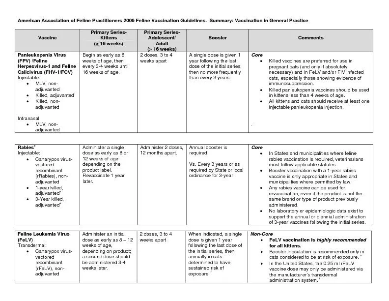
16. PREVENTIONCats that survive an infection develop immunity that likely protects them for the rest of their livesPassive immunity: kittens receive temporary immunity through the transfer of antibodies in the colostrumThere are vaccines that offer the best protection from feline parvovirus infection.Most young kittens receive their first vaccination between six and eight weeks of age and follow-up vaccines are given until the kitten is around 16 weeks of age. Adult vaccination schedules vary with the age and health of the cat, as well as the risk of FP in the area.
17. BIBLIOGRAPHYhttps://www.avma.org/public/PetCare/Pages/feline-panleukopenia.aspxhttps://www.sciencedirect.com/topics/veterinary-science-and-veterinary-medicine/feline-panleukopeniahttp://www.abcdcatsvets.org/abcd-guidelines-on-feline-panleukopenia-2012-edition/https://www.sciencedirect.com/topics/neuroscience/feline-panleukopenia-virus