
Myeloid Lymphoid Histiocytic Myeloid neoplasms 1 Myeloproliferative neoplasms 2 Myelodysplastic syndromes 3 Acute myeloid leukemia Features Recurrent genetic mutations Increased bone marrow cellularity ID: 1045392
Download Presentation The PPT/PDF document "Hematopoietic malignancies" is the property of its rightful owner. Permission is granted to download and print the materials on this web site for personal, non-commercial use only, and to display it on your personal computer provided you do not modify the materials and that you retain all copyright notices contained in the materials. By downloading content from our website, you accept the terms of this agreement.


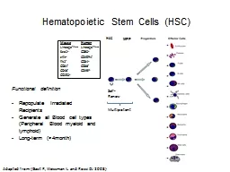

1. Hematopoietic malignanciesMyeloidLymphoidHistiocytic
2. Myeloid neoplasms(1) Myeloproliferative neoplasms(2) Myelodysplastic syndromes(3) Acute myeloid leukemiaFeatures:Recurrent genetic mutationsIncreased bone marrow cellularityTendency to progress to AMLRisk factors:Chemicals (benzen, pesticides), radiation, congenital diseases (Fanconi), smoking, PNH
3. Normal Adult cell count in BM aspirate smearBlasts: <5%Monocytes: <5%M:E 3-4Plasma cells: <3%
4. Aspirate smear from bone marrow shows normal myelogenesis and erythropoiesis
5. Normal bone marrow cellularity depends on age (100-age)%
6. Myeloproliferative neoplasmsChronic disorders hyperproliferation of neoplastic myeloid progenitors that retain the capacity for terminal differentiationMutation in tyrosine kinase proteins which results in permanent activation of growth factorsPersistent peripheral blood cytosis (one or more lines)The neoplastic progenitors tend to seed secondary hematopoietic organs (the spleen, liver, and lymph nodes), resulting in hepatosplenomegaly (caused by neoplastic extramedullary hematopoiesis)
7. Chronic myelogenous leukemiaa balanced (9;22) translocation that moves ABL from chromosome 9 to a position on chromosome 22 adjacent to BCRThe new chr22 is known as Philadelphia chromosomeThe BCR-ABL fusion gene has a tyrosine kinase activity, stimulating the proliferation and prolonged survival of granulocytic and megakaryocytic cells
8.
9. manifestationsPeripheral blood shows markedly increased WBC count, sometimes exceeding 100,000 cell/uLMost of the cells are neutrophils, metamyelocytes and myelocytesBasophils and eosinophils are also increasedThrombocytosis and anemia are commonThe bone marrow is hypercellular owing to increased numbers of granulocytic and megakaryocytic precursorsSpleen is enlarged with extramedullary hematopoiesis
10. Chronic myelogenous leukemia-peripheral blood smear. Granulocytic forms at various stages of differentiation are present
11. Primary MyelofibrosisBrief period of granulopoiesis and megakaryopoiesis, rapidly followed my BM fibrosis and elimination of hematopoietic elementsThe fibroblast proliferation is stimulated by platelet-derived growth factor and transforming growth factor β released from neoplastic megakaryocytesHematopoiesis takes place in spleen and liverRBC’s escaping the fibrotic stroma in BM are deformed and take the shape of “tear-drops”
12. ManifestationsBM is initially hypercellular with increased atypical megakaryocytesPB: leukocytosis, shift to left, thrombocytosis, anemia, nucleated RBCs, tear drop cellsLater in disease, become fibrotic and hypocellular, pancytopeniaSpleen shows marked extramedullary hematopoiesis
13. Two nucleated erythroid precursors and several teardrop-shaped red cells are evident
14. BM biopsy shows hypocellular marrow, spindle shaped stroma and atypia of megakaryocytes
15. Essential ThrombocythemiaChronic MPN involves primarily megakaryocytesSustained thrombocytosis (>450x109/L)Increased number of large mature megsTendency for thrombosis and hemorrhage+ Jak2 in 50%No BM fibrosis+/- splenomegaly
16. ET: left: increased number of megakaryocytes. Right: megakaryocytes are large, mature with hyperlobated nuclei
17. Myelodysplastic syndromesGroup of clonal stem cell disorders characterized by maturation defects that are associated with ineffective hematopoiesisHematopoietic cells are morphologically abnormal, stay within the bone marrow and hence the patients have peripheral blood cytopenias The hallmark of MDS is persistent (refractory) peripheral cytopenia and BM morphologic dysplasia
18. PathogenesisCytogenetic analysis commonly reveals chromosomal aberrationsPrimary (idiopathic): more common, risk factors? Secondary (therapy related): History of chemotherapy or radiotherapy 2-8 years ago All forms of MDS can transform to AML, but transformation occurs with highest frequency and most rapidly in t-MDS
19. FindingsErythroid: megaloblastoid nuclei, nuclear/cytoplasmic asynchrony, multinucleation, ring sideroblastsGranulocytes: hyposegmented nuclei, hypogranular cytoplasmMegakaryocytes: small size, hypolobated nucleiDysplasia can occur in a single or multiple linesWith time, blast increases (5-19%), and can progress to acute leukemia
20. Upper left: ring sideroblasts (iron stain), upper right: dysplastic erythroid precursor (multinucleation), lower right: dysplastic mega (small, hypolobated), middle: dysplastic neutrophil (hypogranulated cytoplasm, hyposegmented nucleus)