
Prepared by Kurt Schaberg MD Germ Cell Tumors 3 main subtypes depending on age and if they are derived from germ cell neoplasia in situ GCNIS Most common Germ cell tumors derived from GCNIS Pos ID: 958622
Download Pdf The PPT/PDF document "Testicular Tumors" is the property of its rightful owner. Permission is granted to download and print the materials on this web site for personal, non-commercial use only, and to display it on your personal computer provided you do not modify the materials and that you retain all copyright notices contained in the materials. By downloading content from our website, you accept the terms of this agreement.


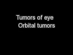

Testicular Tumors Prepared by Kurt Schaberg MD Germ Cell Tumors 3 main subtypes depending on age and if they are derived from germ cell neoplasia in situ (GCNIS ). Most common: Germ cell tumors derived from GCNIS (Post - pubertal type ) (Type 2, below) , which is often sub - grouped into seminoma and non - seminoma germ cell tumors Although only 1% of all male cancers, they are the most common cancers among young men between puberty and 40s. Risk factors: Family history, cryptorchidism, subfertility, pesticides, marijuana, microlithiasis. Although can be aggressive tumors, with current treatments can often be cured as very responsive to chemoradiation. Last updated: 10/12/2020 Germ Cell Neoplasia In situ (âGCNISâ) Proliferation of atypical germ cells within seminiferous tubules Large , angulated nuclei with coarse chromatin (resemble seminoma cells) Often located at base of tubules with prominent halos Often present in nearby parenchyma adjacent to most associated germ cell tumors. Often absent spermatogenesis. Identical IHC profile to seminoma: OCT3/4, cKit (+) Precursor lesion: ~50% progresses to overt Germ Cell Tumor within 5yrs. Intermediate stages between GCNIS and invasion: Intratubular seminoma â complete filling of expanded seminiferous tubule by neoplastic cells with obliteration of normal components. Type Tumors Age Derived from GCNIS Genotype Behavior 1 Teratoma (prepu
bertal) Yolk sac tumor (prepubertal) Dermoid cyst Usually 6 yrs No Diploid or aneuploid. No i12p gains Very good. Mostly benign. 2 Seminoma Embryonal carcinoma Choriocarcinoma Yolk sac tumor Mixed Germ cell tumor Post - pubertal Usually 20s - 30s Yes Aneuploid Frequent gains and losses. Overexpression of isochrome 12p Malignant, but responsive to therapy 3 Spermatocytic Tumor Usually � 50 yrs No Aneuploid No i12p gains Excellent Intratubular non - seminoma â Same concept, but almost exclusively embryonal carcinoma. Thought to be reprogrammed GCNIS cells. Embryonal Carcinoma Seminoma Choriocarcinoma Most common germ cell tumor (~50%). Present with mass. Usually unilateral. Grossly solid, fleshy, lobulated, cream - colored. Large polygonal cell s with clear to eosinophilic cytoplasm (full of glycogen), distinct cell membranes , vesicular chromatin, and prominent nucleoli Fibrous septae and nested architecture Lymphocytic infiltrate ; Sometimes granulomas . GCNIS usually in residual tubules. Rare syncytiotrophoblasts. IHC: (+) OCT3/4, CD117, D2 - 40, SALL4 Elevated serum LDH, rarely hCG . First site of metastases often retroperitoneal lymph nodes . Molecular: majority have isochrome 12p ; ckit mutations in many. Prognosis: Good if treated. Think: Clear/White color Second - most common testicular GCT. Usually part of a mixed GCT Rudimentary epithelial differentiation Large, cro
wded, âPrimitiveâ pleomorphic cells Vesicular nuclei with prominent nucleoli. Coarse, basophilic chromatin. Amphophilic cytoplasm. Variable architecture (nests, sheets, papillae, glands) Prominent mitoses and apoptotic bodies. IHC: (+)CD30, OCT3/4, AE1/AE3, SALL4 Molecular: Isochrome 12p amplification Aggressive, but often responds to chemotherapy Think: Purple color Usually part of a mixed GCT. Malignant cytotrophoblasts and trophoblasts (mononuclear with light cytoplasm ) and syncytiotrophoblasts (multinucleated with deeply eosinophilic cytoplasm ) . Abundant Hemorrhage IHC: (+) hCG . Syncytiotrophoblasts: (+) inhibin, glypican - 3. Cytotrophoblasts: (+) SALL4, p63, GATA3 Very elevated Serum hCG â ( similar to LH and TSH) â gynecomastia, thyrotoxicosis Most aggressive GCT . Frequent hemorrhagic metastases. Less responsive to treatment. Think: Red color Yolk Sac Tumor, Postpubertal - type Teratoma, Postpubertal - type akaÍ âEndodermal Sinus Tumorâ or âYSTâ Almost always a component of mixed GCT Many patterns/architecture (often combined) Most common = reticular/microcystic (Honeycomb meshwork) Can also be solid, papillary, glandular, etc Í Often hypocellular myxoid areas Schiller - Duval Bodies (endodermal sinus)( â ) Refractile eosinophilic hyaline globules ( â ) Band - like intercellular basement membrane material Can have âhepatoidâ areas resembling liver tha
t stains with liver markers. IHC: (+)AFP, Glypican - 3, SALL4, AE1/AE3, Elevated Serum AFP Post - chemo can get sarcomatoid YST Think: Pink color Composed of tissues from one or more germinal layers . May be composed of differentiated mature tissue or immature, embryonic - type tissue . Often part of a mixed GCT. In contrast to ovary, pretty much all teratomas in postpubertal testis are malignant ! Can see virtually all tissue types including epithelial and mesenchymal. Often multiple cysts lined by glandular or squamous epithelium. Frequent immature neuroectodermal structures. IHC: Differentiated elements express profile of that tissue type. Often areas of cytologic atypia, including primitive mitotically active stroma cuffing glands. If a dysplastic component forms a nodule that is larger than a 4X field (5 mm) â somatic - type malignancy arising in a teratoma. Usually a sarcoma, most commonly rhabdomyosarcoma. Most common component in a treated GCT. Rare situation where can be benign teratoma in an adult : Dermoid cysts, or, organoid morphology with prominent components of ciliated epithelium and smooth muscle and no GCNIS, isochrome 12p, or testicular scarring. Mixed Germ Cell Tumor Regressed Germ Cell Tumors Germ cell tumors that have undergone either partial or complete regression (âburnt - outâ), leaving behind a well - delineated nodular focus of scaring fibrosis in the
testis. Can present with metastatic disease, but primary has completely regressed . Can be seminoma or Non - seminoma. Scar findings: Well - demarcated scar, Coarse calcifications within tubules , chronic inflammation, hyalinized tubular ghosts. Nearby findings: GCNIS , tubular atrophy, microliths Malignant tumors with more than one germ cell tumor component . Clinically regarded as â non - seminoma â (even if seminoma present)Í Majority of all non - seminomatous GCT are mixed. Must report approximate % of each component . NoteÍ Syncytiotrophoblasts â choriocarcinoma (can see in other tumors, like seminoma) Special subtypes: Polyembryoma â combination of embryonal carcinoma and YST resembling an embryo Diffuse embryoma â orderly combination of embryonal carcinoma and YST in parallel flat layers (pictured â ). Spermatocytic tumor Relatively rare. Generally excellent prognosis . NOT associated with GCNIS or cryptorchidism NOT a component of mixed GCT Usually occurs in OLD er men �(50yo) Polymorphous cell population ( 3 cell types : small, medium, and large) Poorly - defined cell membranes. Dense cytoplasm . Round nuclei with dense to granular chromatin. Diffuse to multinodular pattern of growth. Frequent cystic change/ edema . No significant inflammation/granulomas IHC: Negative for usual seminoma markers (e.g., OCT3/4). (+) cKit and SALL4 Can undergo sarcomatous transform
ation. Yolk Sac Tumor, Prepubertal - type Composed of elements resembling somatic tissues from one or more germ cell layers. Primarily occurs in prepubertal males years old (but can see in older) In contrast with Postpubertal - type: Benign behavior . Do not recur or metastasize. NO association with GCNIS or isochrome 12p amplification. NO cytologic atypia. NO association with mixed GCT. As such, they are most akin to the mature cystic teratomas seen in the ovary. Frequently include skin structures, ciliated epithelium, fat, cartilage, bone, and muscle in organoid structures. No significant cytologic atypia. Normal surrounding testicle : No GCNIS, tubular atrophy, scars, microlithiasis, necrosis, or impaired spermatogenesis (which might suggest a GCNIS - derived GCT) Specialized variants: Dermoid Cyst : replicate skin in an organoid arrangement. Squamous epithelium with adnexal structures. Cured by excision. Epidermoid Cyst : Unilocular cyst with squamous lining and keratinaceous debris. No adnexal structures or other elements. Cured by excision. Well - differentiated Neuroendocrine Tumor : Similar morphology to elsewhere. Often pure. Usually good behavior. Only variant that can behave aggressively. Rare. Usually in young boys 6 years old Identical morphology and IHC profile to postpubertal - type. Secretes AFP However, unlike postpubertal YST : NOT associated with GCNIS. NO isochrome 1
2p amplification . Excellent survival , even with advanced stage. Usually pure, but can see in combination: Mixed teratoma and yolk sac tumor, prepubertal - type Teratoma, Prepubertal - type Immunohistochemistry of Germ Cell Tumors GCNIS Seminoma Embryonal Carcinoma Yolk Sac Tumor Choriocarcinoma Teratoma Spermatocytic Tumor Metastatic Carcinoma Other Tumors AE1/AE3 - ± + + + + - + Many! OCT3/4 + + + - - - - - Rare NSCLC and RCC and large cell lymphoma CD30 - - + - - - - ± Lymphomas, melanoma, nasopharyngeal carcinoma, mesenchymal tumors Glypcian - 3 - - - + + ± ± HCC, gastric cancers, syncytiotrophoblasts D2 - 40 + + ± - - ± - ± Gliomas, meningiomas, mesothelial tumors, lymphatic tumors, PLAP + + + ± + - - ± Numerous adenocarcinomas (colon, endometrium, etc..) SALL4 + + + + ± ± + ± Hematologic malignancies, rhabdoid tumors, Wilms tumor, lots of GI adenocarcinomas among others β hCG - - - - + - - ± Other trophoblastic tumors, syncytiotrophoblasts cKIT (CD117) + + - ± - - ± ± Lots of tumors AFP - - ± + - ± - ± Hepatocellular tumors, etc.. CK7 ± ± + - + + ± Many carcinomas Modified from: WHO classification of Tumors of the Urinary System and Male Genital Organs. 4 th ed. Leydig Cell Tumor Sex Cord - Stromal Abundant, eosinophilic granular cytoplasm . Diffuse growth. Uniform round cells. Round, central nuclei with prominent nucleoli. Frequent Reinke crystals ( â ) Usually asymptomatic , but chil
dren can present with precocious puberty as the tumor can secrete steroid hormones (e.g., testosterone). Most common testicular sex cord - stromal tumor. Vast majority are benign. Sertoli Cell Tumor Often shows at least focal tubular differentiation . Usually moderate pale cytoplasm. Rarely diffuse growth. Unique IHC: Frequent nuclear β - catenin , WT - 1, CK AE1/AE3, and neuroendocrine markers. Vast majority are benign. Variant : Sclerosing Sertoli Cell Tumor â extensively hyalinized stroma with cells arranged in tight cords and clusters Rare. More common in kids. Vast majority are benign. A little variable, but often stain with some combination of: Inhibin, calretinin, SF - 1 , FOXL2, Melan A Factors associated with Malignant behavior in Sex Cord - Stromal Tumors: Cytologic Atypia, Abundant Mitoses, Large size, Vascular Invasion, Necrosis, Infiltrative growth (Pretty common - sense bad findings ; - ) Other Tumors Large Sertoli cells with abundant granular eosinophilic cytoplasm Calcifications (focal, psammomatous to large, plaque - like) Often prominent neutrophilic infiltrate . NO nuclear β - catenin Frequently associated with Carney complex Frequent PRKAR1A mutations Intratubular Large Cell Hyalinizing Sertoli Cell Neoplasia: Expanded seminiferous tubules with large Sertoli cells with pale cytoplasm accompanied by prominent basement membrane deposits around and within tubules
( â ) . Almost exclusively in Peutz - Jegherâs syndrome ( think: like SCTATs! ). Often present as prepubertal males with gynecomastia (aromatases made by tumor convert androgens â estrogen). Always benign. Fibroma/Thecoma: Resemble ovarian counterparts. Benign. Rare. Unencapsulated proliferation of spindled cells with scant eosinophilic cytoplasm. Large Cell Calcifying Sertoli Cell Tumor Granulosa Cell Tumors Adult Granulosa Cell Tumors Rare. Often asymptomatic, but can secrete estrogen Cells: Scant pale architecture with grooved nuclei Varied architecture : Sheet - like, trabecular, ribbon - like, microfolicular (with â Call - Exner bodies â filled with pink secretions)Í Molecular: Frequent FOXL2 point mutations Juvenile Granulosa Cell Tumors Rare. Almost all in first decade, often before 6 months old. Usually presents as a mass. Macrofollicles with mucinous secretions Round nuclei with NO GROOVES Similar to the more common ovarian counterpart Gonadoblastoma Hematolymphoid Tumors Most common testicular tumor in men over 50 years old. Can be primary or part of systemic involvement. Often obliterate the seminiferous tubules centrally with peripheral intertubular spread . Diffuse Large B - Cell Lymphoma comprises ~90% of primary testicular lymphomas. Same stains as elsewhere. Miscellaneous Tumors Germ cells resembling GCNIS cells and spermatogonium Sex cord cells resembling immature
granulosa cells Arranged in round nests with round deposits of eosinophilic basement membrane Frequent calcifications Develop in individuals with gonadal dysgenesis . Can progress to a germ cell tumor, often seminoma. Other Tumors Ovarian - type Epithelial Tumors: Resemble entire spectrum of ovarian type epithelial tumors. Most commonly Serous and Mucinous, with most being Serous Borderline Tumors, which do not recur or metastasize. Rete Testis Adenoma: Very rare. Benign tumor of rete epithelium that spans the spectrum from packed tubules (adenoma) to tumors with a cystic component (cystadenoma), papillary architecture (papillary cystadenoma), or glands with prominent fibrous tissue (adenofibroma). Rete Testis Adenocarcinoma: Very Rare. Malignant. Diagnosis of exclusion. Malignant gland forming tumor of rete epithelium. Must be centered in hilum of testis, patient must have no other similar primary, and other Dxâs (eÍgÍ, mesothelioma), must be excluded. Poor prognosis. Myoid Gonadal Stromal Tumor : Spindle cell tumor near rete testis with features of gonadal stroma and smooth muscle. Circumscribed with densely packed spindled cells arranged in short fascicles. IHC: (+)SF1, FOXL2, SMA, S100. Adenomatoid Tumor Mesothelioma Rare. Malignant proliferation of mesothelial cells arising from tunica vaginalis. Mass envelops testicle , often invading it. Like in the pleura, variable appearance. Of
ten epithelioid with papillary or tubulopapillary architecture. Less associated with asbestos. IHC: Usual mesothelial markers (see above) Paratesticular Tumors Benign Mesothelial tumor. Most common neoplasm of paratesticular region Often based in the epididymis and 2cm. Irregularly shaped gland - like microcystic spaces composed of flattened or cuboidal cells with associated fibrous stroma and lymphoid aggregates. Bland cytologic features. Helpful feature : "thread - like bridging strandsâ ( â ) Sometimes signet ring - like vacuolated cells. Solitary, localized. IHC: Mesothelial markers: D2 - 40, Calretinin, WT - 1, CK5/6 , CK AE1/AE3. Papillary Cystadenoma of the Epididymis Rare. Benign Tumor of Epididymal ducts. Associated with von Hippel - Lindau syndrome (but is more often sporadic) Cystic structures that are focally papillary. Clear columnar/cuboidal cells with abundant clear cytoplasm . Frequent reverse nuclear polarity. Colloid - like secretions. Morphologically (and immunophenotypically) resembles papillary clear cell RCC (see separate Kidney guide) Adipocytic Tumors Skeletal Muscle Tumors Smooth Muscle Tumors Lipoma: Benign. Most common mesenchymal tumor of region. Consist of entirely mature adipocytes. Well - differentiated Liposarcoma: Common paratesticular sarcomaÍ Recur, but wonât metastasize unless dedifferentiate. Varying proportion of adipocytes, fibrous bands with enla
rged, hyperchromatic stromal cells, and occasional lipoblasts. Can see inflammation/myxoid change. Giant marker and/or ring chromosomes â MDM2 amplification . If large or questionable atypia â consider getting FISH to help differentiate. Leiomyoma: Benign. Somewhat common. Consist of entirely bland smooth muscle (like in other locations). Leiomyosarcoma: Common paratesticular sarcoma. Fascicles of spindled cells with brightly eosinophilic cytoplasm and âcigar - likeâ blunt nucleiÍ Significant atypia, mitoses, and/or tumor necrosis. IHC: Desmin, SMA, H - Caldesmon , Calponin Rhabdomyoma: Benign. Very rare. Most often adolescents. Rhabdomyosarcoma: Often in children or young adults and Embryonal subtype with primitive round or spindled cells and variable eosinophilic rhabdomyoblasts with abundant eccentric cytoplasm. Often good prognosis. IHC: MyoD1, Myogenin , Desmin. No Algorithmic Diagnosis of Testicular Tumors Clear Cytoplasm: Glandular and/or Tubular growth: Seminoma [Oct3/4+, CD30 - , Glypican3 - ] Embryonal [Oct3/4+, CD30+, Glypican3 - ] Yolk Sac Tumor [Oct3/4 - , CD30 - , Glypican3+, AFP+] Lymphoma [CD45+, Oct3/4 - , inhibin - ] Sertoli Cell Tumor [Inhibin+, SF1+, Nuclear β - catenin] Spermatocytic Tumor [Oct3/4 - , CD30, Glypican3 - ] Metastatic Adenocarcinoma [CK+, SALL4 - , Oct3/4 - , CD30 - , Glypican3 - ] GCNIS Present? Nests p olygonal cells arranged wit
h fibrous septae with lymphocytes? Predominantly Intertubular Growth? Large, irregular partially clear nuclei? Focal Glandular Architecture? 3 Distinct Cell Types? Start Yes Yes Yes Yes Yes Yes No No No No No Seminoma [Oct3/4+, CD30 - , Glypican3 - ] Embryonal [Oct3/4+, CD30+, Glypican3 - ] Yolk Sac Tumor [Oct3/4 - , CD30 - , Glypican3+, AFP+] Sertoli Cell Tumor [Inhibin+, SF1+, Nuclear β - catenin] Rete Testis Neoplasm [centered in hilum, CK+, PAX8 - , CEA+, EMA+] Metastatic Adenocarcinoma [CK+, SALL4 - , Oct3/4 - , CD30 - , Glypican3 - ] GCNIS Present? Cells with large crowded, irregular nuclei lining gland - like spaces? Predominantly intertubular and intralymphatic growth?? Variably sized cells, hyaline globules, variable patterns? Tubular growth with solid tubules? Start Yes No Yes Yes No Yes No No Yes No Algorithms modified from: Urologic Surgical Pathology. Liang Chen et al. 2020. Microcystic: Pink Cells with Abundant cytoplasm (Oxyphil): Well - differentiated Neuroendocrine Tumor [CK+, Synaptophysin+, Inhibin - ] Leydig Cell Tumor [Inhibin+] Large Cell Calcifying Sertoli Cell Tumor [Inhibin+] Consider: Metastatic Carcinoma, Melanoma, Hematolymphoid tumors Hepatoid Yolk Sac Tumor [CK+, Glypican3+, AFP+, Oct3/4, CD30 - , Inhibin - ] Adenomatoid Tumor [CK+, Calretinin - ] Round nuclei with prominent nucleoli, ± lipofuscin or Reinke Crystals? Insular and trabec
ular patterns, coarse chromatin, ± teratomatous elements? Fibromyxoid stroma with neutrophils and calcifications? Highly pleomorphic nuclei, Prominent intratubular growth? Presence of other patterns, Lots of mitoses, and associated GCNIS? Start Yes No Yes Yes No Yes No No Yes No Seminoma [Oct3/4+, CD30 - , Glypican3 - , CK - ] Leydig Cell Tumor: Oct3/4+, CD30+, Glypican3 - Adenomatoid Tumor: CK+, Calretinin+ Yolk Sac Tumor ( post - pubertal ) [CK+, Oct3/4 - , CD30 - , Glypican3+, AFP+, PLAP+] Sertoli Cell Tumor [Inhibin+, SF1+, Nuclear β - catenin] Juvenile Granulosa Cell Tumor [Inhibin+, AFP - , Glypican3 - ] GCNIS Present? Variably sized nuclei and flattened nuclei linin cysts? Prominent cords of tumor cells, Lipid rich cells, and hyalinized stroma? Abundant eosinophilic cytoplasm? Neonate? Start Yes No Yes Yes No Yes No No Yes No Yolk Sac Tumor ( pre - pubertal ) [CK+, Oct3/4 - , CD30 - , Glypican3+, AFP+, PLAP+] Spindle Cells: MPNST, Melanoma, Liposarcoma [S100+ at least partial] Sarcomatoid Carcinoma or Mesothelioma [CK+] Fibrothecoma [SMA - , S100 - ] Leiomyosarcoma, Rhabdomyosarcoma [Desmin+] Round nuclei with prominent nucleoli, ± lipofuscin or Reinke Crystals? Bland Nuclear Cytology with no Mitoses? Keratin, S100, Desmin staining? Start Yes No Yes No Leydig Cell Tumor [Inhibin+] Myoid Gonadal Stromal Tumor [SMA+, S100+] Smooth Muscle Actin and S100 stai