
Syndrome spotter Definition Resources Grouped by aetiology Grouped by E ar N ose T hroat Syndrome spotter Definition a collection of traits health problems and ID: 944313
Download Pdf The PPT/PDF document "Syndrome spotter" is the property of its rightful owner. Permission is granted to download and print the materials on this web site for personal, non-commercial use only, and to display it on your personal computer provided you do not modify the materials and that you retain all copyright notices contained in the materials. By downloading content from our website, you accept the terms of this agreement.


Syndrome spotter Syndrome spotter ⢠Definition ⢠Resources ⢠Grouped by aetiology ⢠Grouped by â E ar â N ose â T hroat Syndrome spotter ⢠Definition â âa collection of traits, health problems, and/or birth defects in an individual which usually has a single underlying causeâ â eg gene defect such as Downs â Syn + Dramien âto run togetherâ â Sequence Single cause with multiple cascading effects â Eg Pierre Robin â Association recognizable pattern without single cause (yet!) â Eg CHARGE, VATERS ⢠Resources Syndrome spotter ⢠Definition ⢠Resources â Smiths - recognisable patte
rns of human malformation â London dysmorphology database www.lm databases .com â Medline search â Google search â www.emedicine.com â European Surveillance of Congenital Abnormalities http://www.eurocat.ulster.ac.uk/pdf/Ester/EUROCAT%20Guide%206%20Version%203.pdf Grouped by syndrome ⢠Chromosomal; Downs, Turners, Cri du Chat ⢠Craniofacial: Crouzons, Aperts, Pfeifers ⢠Palatal; Clefts, Pierre Robin, VCF ⢠Connective tissue; MPS, EB; OI; ⢠Branchial arch; Goldenhars ⢠Obesity; Beckwith Weiderman, Prader Willi ⢠Fetal: Alcohol, warfarin, rubella ⢠Others Treacher -
collins, CHARGE Grouped by site ⢠Ear â Abnormal pinnae eg: Treacher Collins â Deafness eg: Wardenburgs ⢠Nose â Abnormal external nose eg:Cranio metaphyseal dysplasia, Hallerman Streif â Choanal atresia eg: CHARGE ⢠Throat â Palate eg: Pierre Robin, VCF â Airway obstruction eg: Prader - Willi Listed alphabetically ⢠Achondroplasia ⢠Alports ⢠Alstromâs ⢠Aperts ⢠Beckwith Wiedermann ⢠Brancho oto renal ⢠Campomyelic Dysplasia ⢠Cleft syndromes ⢠CHARGE ⢠Chondrodysplasia Punctata ⢠CMV Infection ⢠Congenital Rubella â¢
Cornelia de Lange ⢠Craniometaphyseal dysplasia ⢠Cri du Chat ⢠Crouzons ⢠Di George ⢠Downs ⢠EB ⢠EEC ⢠Fetal alcohol ⢠Fetal Warfarin ⢠Fraserâs ⢠Goldenharâs ⢠Gorlinâs ⢠Hallerman Streif ⢠Holoprosencephaly ⢠Jervell ⢠Klippel Fiel ⢠Larsens ⢠McUne Albright ⢠Moebius ⢠MPS ⢠NF ½ ⢠boonanâs ⢠Osteogenesis Imperfecta ⢠Opiz G Osteopetrosis Pallitser Hall Pendreds Pierre Robin Prader Willi Pfeiffers Refsumâs Saethre Chotzen Stickler Toxoplasmosis Treacher Collins Turners Usherâs VATERS VCF Waardenburgâs Chromosomal Anomalies
- Downs ⢠Trisomy 21 â Translocation 2% â Mosaicism 2% ⢠1866, John Langdon Down ⢠1:800 to 1,000 live births. ⢠increased risk in women aged over 35 â 35 year old 1: 400 â 40 year old 1: 110 â 45 year old 1: 35. ⢠Antenatal diagnosis â CVS â Nuchal thickness âThe face is flat and broad, and destitute of prominence. The cheeks are roundish, and extended laterally. The eyes are obliquely placed, and the internal canthi more than normally distant from one another. The palpebral fissure is very narrow. The forehead is wrinkled transversely, from the constant assistance which the levatores pebrarum deriv
e from the occipito - frontalis muscle in the opening of the eyes. The lips are large and thick, with transverse fissures. The tongue is long, thick and is much roughened. The nose is small. The skin has a slight dirty - yellowish tinge, and is deficient in elasticity, giving the appearance of being too large of the body â Chromosomal Anomalies - Downs ⢠Women with Downs have 50 percent chance that their child will have Down syndrome. ⢠Men with Down syndrome are believed to be sterile. Chromosomal Anomalies - Downs ⢠General Features â Simian crease, a single deep crease across the center of the palm â Oblique palpebral fissures
â Muscle hypotonia â Hyperflexibility â Most of these features also occur in general population Chromosomal Anomalies - Downs â Epicanthal folds small skin folds on the inner corner of the eyes â Flat facial profile ⢠depressed nasal bridge ⢠small nose â Cardiac anomalies â IQ - variable â Low immunity â Unstable neck ⢠Atlanto - occipital joint 15% ⢠PM:2527213 â Subglottic stenosis Chromosomal Anomalies - Downs ⢠ENT features http://www.emedicine.com/ped/topic615.htm â External ears ⢠Small canals â Middle ears ⢠Increased incidence of glue ear (age related) ⢠Otoa
coustic emissions often non - reproducible, even in patients with normal hearing abilities ⢠? Abnormal ossicles â Mixed deafness 7% â Sensorineural deafness 8% â Airway obstruction from small PNS with large tonsils and adenoids â Macroglossia Chromosomal Anomalies ⢠Abnormal 5p â cri du chat â Partial atresia larynx â http://www.emedicine.com/ped/topic504.htm Chromosomal Anomalies - Turners ⢠General â XO â Short stature â Ovarian dysgenesis â Variable IQ â Increased carrying angle ⢠ENT features â Increased incidence glue ear â Sensorineural deafness â http://www.emedicine.com/ped/to
pic2330.htm Craniofacial Syndromes ⢠Crouzons - craniosynostosis . http://www.emedicine.com/ped/topic511.htm ⢠Aperts - with syndactyly ⢠Pfeiffers - broad short thumbs - oversized toes Craniofacial Syndromes - Aperts ⢠First described 1906 ⢠Single gene Mutation chromosome 10q ⢠Spontaneous 1:120,000 ⢠Autosomal dominant pattern ⢠http://www.emedicine.com/ped/topic122.htm Craniofacial Syndromes - Aperts Craniosynostosis with syndactyly ⢠General features â Craniosynostosis â Syndactyly â Hypertelorism â Proptosis ⢠ENT features â Airway o
bstruction â Abnormal ear â Hearing loss â Complete cartilage rings Craniofacial syndromes â Hearing loss ⢠Pinna abnormalities include low set, small or posteriorly rotated ears, such as those seen in our Apert's, Pfeiffer's and Saethre - Chotzen patients. A prominent antihelical fold is frequently seen in patients with Saethre - Chotzen. ⢠External canal atresia is also a feature of craniosynostosis syndromes. It was found in 13% of patients with Crouzon's syndrome and is also reported to be quite common with Pfeiffer's syndrome. ⢠Middle ear abnormalities consist of both congenital ossicular fixation and eustachian tube dysfunct
ion. â Ossicular fixation is also a feature of other craniosynostosis syndromes, including Crouzon's, in which lateral chain abnormalities may be found. â Stapes fixation is the most classically described malformation in patients with Apert's syndrome. Bergstrom reported a perilymph gusher during stapedectomy in one patient operated on for stapes fixation. Because of this complication and the frequency of middle ear effusion and infection, the performance of stapedectomy has been condemned by a number of authors, while other simply warn that it should be approached with caution. â Eustachian tube dysfunction leading to effusions, perforation, and ch
olesteatoma is thought to be secondary to altered nasopharyngeal and skull base anatomy and is extremely common. Significantly, as these patients grow, the relative hypoplasia of the skull base is often accentuated and middle ear pathology frequently persists into adulthood. ⢠Inner ear pathology is poorly characterized in these patients, although there is an increased incidence of sensorineural hearing loss. Craniofacial Syndromes - Pfeiffers Craniosynostosis with short broad thumbs ⢠Craniosynostosis syndrome resulting from premature fusion of the sutures of the skull resulting in skull deformity. ⢠Abnormal skull growth, which results in a
pointed or conical head, is also responsible for underdevelopment of the mid - face (upper jaw bone), high arched palate and prominent lower jaw are characteristic. ⢠Eyes are wide set and bulge. ⢠Teeth erupt in improper positions. ⢠Mild hearing loss due to a defect in the middle ear may be present. ⢠Thumbs are short and broad; toes are oversized. ⢠Hands and feet may be webbed. ⢠Autosomal dominant ⢠http://www.emedicine.com/derm/topic325.htm Cleft syndromes Cleft syndromes Cleft syndromes Velo cardio facial syndrome - General features ⢠Palate (Velo - ) â Overt, submucous or occult cleft palate â Cleft lip
(uncommon) ⢠Heart (Cardio - ) â VSD â ASD â Pulm atresia â Teratology of fallot â Vascular ring ⢠Orofacial â Retrognathia â Assymetric face â Teeth ⢠Missing ⢠Enamel hypoplasia ⢠Others â Learning difficulties â Feeding difficulties â Immune deficiency ⢠Test â (FISH). fluorescent in situ hybridization â FISH is a type of specialized chromosome analysis Velo cardio facial syndrome - ENT features ⢠Ears â Over folded helix â Attached lobules â Protruberant cup ears â Microtia â AOM â Glue Ear â SN loss â Narrow canals â http://www.emedicine.com/ped/
topic2395.htm ⢠Nasal â Prominent bulbous nasal tip â Narrow nares â Upper airway obstruction â Small adenoids ⢠Laryngeal â Anterior laryngeal web â Laryngomalacia Connective tissue - Mucopolysaccharidoses ⢠General features â Coarse or rough facial features â Thick lips, enlarged mouth and tongue) â Dwarfism â Skeletal irregularities â Visceromegaly â Short claw - like hands, â Progressive joint stiffness â Recurring respiratory infections â Heart disease. ⢠http://healthlibrary.stanford.edu/resources/inte rnet/bodysystems/metabolic_m.html ⢠ENT features â Obstructive airway d
isease and obstructive sleep apnea. ⢠Diffuse infiltration around airway â Hearing loss (see below) Mucopolysaccharidoses (MPS) and Mucolipidoses (ML) are genetic lysosomal storage disorders caused by the bodyâs inability to produce specific enzymes. The missing enzyme prevents the normal breakdown and recycling of cells resulting in the storage of these cell deposits in virtually every cell of the body MPS ⢠3 main types of type 1 MPS â Type HS Hurler - Scheie syndrome, ⢠MPS type I H/S produces clinical features that are intermediate between types IH and IS ⢠Type I H/S is milder than type IH and progresses more slowly ⢠Childr
en with this syndrome are usually healthy at birth, with onset of symptoms when aged 3 - 8 years ⢠Survival to adulthood is common ⢠Patients with type I H/S characteristically have corneal clouding, joint stiffness, dysotosis multiplex, and heart disease â cs â Type IH (Hurler Syndrome) â Type IS (Scheie Syndrome). ⢠Mucopolysaccharidosis II (MPS II) â X - linked disorder, which also has the eponym Hunter syndrome, results from the deficiency of iduronate 2 - sulfatase ⢠MPS type III, or Sanfilippo syndrome â The four subgroups of MPS III are as follows: type IIIA (Sanfilippo A), type IIIB (Sanfilippo B), type IIIC (Sanfilippo C),
a nd type IIID (Sanfilippo ⢠Morquio â Husler coined the term dysostosis multiplex to describe the constellation of skeletal findings specific to patients with MPS and other lysosomal storage disorders. These included a large skull with a J - shaped sella, anterior hypoplasia of the thoracic and lumbar vertebral bodies, hypoplasia of the pelvis with small femoral heads and coxa valga, oar - shaped ribs (narrow at the vertebrae and widening anteriorly), diaphyseal and metaphysea l expansion of long bones with cortical thinning, and tapering of the proximal phalanges. ⢠MPS VI â progressive connective tissue organ involvement, resulting from continuo
us storage of dermatan sulfate in the skeleton, heart va lves, spleen, liver, and cornea. â Patients appear healthy at birth and have accelerated growth in the first year, followed by deceleration and short stature la ter in childhood. The diagnosis usually is made in early childhood when organomegaly, corneal clouding, coarse features, enlarged tongue, and joint stiffness al l are apparent. Other complications include hearing loss, chronic respiratory infections, and cardiac insufficiency due to valvular disease. ⢠MPS VII â In severe cases, the condition presents as hydrops fetalis. Neonatal jaundice may be present at birth. More typically, the cl in
i cal features of the disorder become evident in the first few years of life. These early symptoms include dysostosis multiplex with dislocated hips, joint con tractures, and thoracolumbar kyphoscoliosis. In many patients, a J - shaped turcica and odontoid hypoplasia also may occur. Radiographs demonstrate a flattenin g of the vertebral bodies termed platyspondyly. ⢠Many individuals have hearing loss, either conductive (in which pressure behind the ear drum causes fluid from the lining of the middle ear to build up and eventually congeal) or sensorineural. Connective tissue EB ⢠EB simplex is within the epidermis. ⢠Junctional EB is seen at the
level of the lamina lucida within the basement membrane zone. ⢠Dystrophic EB or dermolytic EB is a scarring form of EB which occurs in the deeper tissue at the level the lamina densa or upper dermis. EB - Laryngeal involvement by type http://www.emedicine.com/DERM/topic124.htm Osteogenesis Imperfecta ⢠Type I â Commonest form â Autosomal dominant â Cells from individuals with type I OI secrete about half the normal amount of type I procollagen â In some families dentinogenesis imperfecta is a feature. â Blue sclerae â Tendency to fractures of the long bones, although healing occurs without deformity. â Hearing l
oss â Stapedectomy results ⢠PM:10962673 ⢠PM:15612382 ⢠Operative findings included fixation or thickening of the stapes footplate with normal superstructure configuration and hypervascularization of the promontory mucosa. ⢠http://www.emedicine.com/PED/topic1674.htm Goldenhars, Auriculovertebral ⢠Facial asymmetry ⢠Microtia ⢠Epibulbar dermoid ⢠Vertebral defects ⢠Without eye and vertebral anomolies = hemifacial microsomia ⢠http://www.whonamedit.com/synd.cfm/2300.html Branchio - oto - renal syndrome ⢠Malformations of the outer, middle, and inner ear â conductive, sensorineural, or mixed hearing
impairment ⢠Branchial fistulae and cysts; ⢠Renal malformations, â mild renal hypoplasia to bilateral renal agenesis. ⢠EYA1 gene mutations in approximately 40% ⢠Autosomal dominant. ⢠Extreme clinical variability can be observed in the same family. ⢠Prenatal testing is clinically available. ⢠http://www.boystownhospital.org/Hearing/info/genetics/syndromes/bor.asp Obesity â Prader Willi Morbid obesity with developmental delay, hypogonadism and dwarfism ⢠A disorder of chromosome 15 ⢠Hypothalamic dysfunction: â Hyperphagia â Morbid obesity â Hypogonadism â Cognitive impairment â Difficu
lt behaviour â Growth failure â Hypotonia ⢠Due to a few recent fatalities reported in individuals with PWS who were on growth hormone therapy (GH) some physicians have also added this as an additional risk factor. One possibility (that is currently unproven) is that GH could increase the growth of lymphoid tissue in the airway thus worsening already existing hypoventilation or OSA. Nonetheless, it must be emphasized that there are currently no definitive data demonstrating GH causes or worsens sleep disordered breathing. ⢠http://www.prader - willi.org/Prader - Willi%20Overview.htm Obesity; Beckwith - Wiedemann Syndrome Morbid obesity
with macroglossia ⢠Overgrowth disorder. â First recognized in 1963 â Usually sporadic, occ AD â Risk of hypoglycemia â Incidence 1:15,000 births. â Macroglossia â Abdominal Wall Defects â Increased Growth - Birth Weight and Length usually above average. - Visceromegaly: - Hemihypertrophy: â Typical facial features - Earlobe creases : or pits behind the upper ear. - Prominent occiput: enlarged back of the skull. - Nevus Flammeus: a strawberry mark commonly found on the forehead and eyelids . ⢠http://www.bws - support.org.uk/html/what_is_bws.html Foetal Alcohol Abnormal face with hearing loss
⢠Short palpebral fissues, abnormal philtrum, thin upper lip, hypoplastic midface ⢠Neurodevelopment disorder (more than one may be identified, but not all conditions must be present) ⢠Head circumference 10th percentile ⢠Intellectual impairment ⢠Memory problems ⢠Delayed development ⢠Attachment concerns ⢠Attention deficit disorder ⢠Impaired motor skills ⢠Hyperactivity ⢠Neurosensory hearing loss ⢠Problems with reasoning and judgment ⢠Learning disabilities ⢠Inability to appreciate consequences ⢠Impaired visual/spatial skills ⢠http://www.emedicine.com/ped/topic767.htm Fetal warfarin
⢠Head and neck: â Microcephaly and midfacial hypoplasia. â Eyes: Optic atrophy, corneal opacity, and cataracts. â Nose: Hypoplastic nose with low nasal bridge and choanal atresia. ⢠Thorax: Widely spaced nipples. ⢠Hand and foot: Hypoplasia and shortening of digits. ⢠Muscles: Hypotonia.Bones and joints: ⢠Punctate epiphyses, calcification disorders with stippling of the epiphyses, and bone defects similar to those in chondrodysplasia punctata. ⢠Nervous system: â Brain agenesis, hydrocephalus, agenesis of corpus callosum, meningoencephalocele, and seizures. ⢠Cardiovascular system: â Patent ductus arteriosus, pulmonic s
tenosis, transposition of the great vessels, and anomalous pulmonary veins. ⢠Respiratory system: Airway obstruction. ⢠Growth and development: â Growth and mental retardation. â Behaviour and performance: â Deafness, â Feeding difficulty, and failure to thrive . ⢠http://www.emedicine.com/emerg/topic872.htm Congenital Rubella ⢠Sensorineural deafness, which can progress after birth; ⢠Opthalmic defects such as cataracts; ⢠Cardiovascular defects; ⢠Brain damage, that only occurs after infection between the 3rd and 16th week of gestation, causing mild to severe mental retardation with microcephaly and spastic diplegia
⢠Major structural malformations are rare. ⢠In France, systematic vaccination of male and female newborns was introduced in 1985 and induced a marked reduction in the incidence of CRS (from 13 to 5 cases in 100,000 livebirths). ⢠http://www.emedicine.com/emerg/topic388.htm Treacher Collins syndrome ⢠Downslanting eyes ⢠Notches of the lower eyelids, most frequently on the lateral 1/3 ⢠Mandibular hyploplasia ⢠Prominent nose ⢠Broad mouth ⢠Small chin with a steep angle of the lower jaw ⢠Microtia ⢠Sideburns ⢠Hearing loss, usually conductive. ⢠The nasopharynx may be narrow. ⢠Also â Cleft lip, with
or without cleft palate â Heart defects â Strabismus ⢠http://www.emedicine.com/ped/topic1364.htm CHARGE ⢠Location 2p14? ;7q21;2q33? ⢠Coloboma (iris or retina) ⢠Heart (any defect) ⢠Atresia Choanae ⢠Retardation (growth and development) ⢠Genital (hypoplasia) ⢠Ear (microtia) ⢠(protocol for pts with Choanal atresia) ⢠http://www.emedicine.com/ped/topic367.htm 1 . CT scan after Ephedrine nasal drops (0.5%), suction and vasoconstrict 2. ECHO/Cardiology pre - op 3. Renal ultrasound 4. Ophthalmology 5. Audiology Cornelia De lange ⢠Low - pitched weak cry ⢠Initial hyper
tonicity (100%) ⢠Respiratory and feeding difficulties in newborn period and infancy (71%) â failure to thrive. â gastroesophageal reflux â Developmental delay and mental retardation â Severe speech delay is typical. â mild - to - moderate mental retardation. ⢠Behavioral manifestations â Hyperactivity. â diminished ability to relate. ⢠Ophthalmologic manifestations (50%) ⢠Short stature ⢠Microcephaly (98%) ⢠Facial features â Confluent eyebrows â Long curly eyelashes â Low anterior and posterior hairline â Underdeveloped orbital arches â Neat, well - defined, and arched eyebrows â Long phil
trum â Anteverted nares â Down - turned angles of the mouth â Thin lip (especially upper vermillion border) â Low - set ears â Depressed nasal bridge â High arched palate â Late eruption of widely spaced teeth â Micrognathia â Short neck Abnormal face, feeding difficulties and reflux http://www.emedicine.com/ped/topic482.htm Hallerman Streiff Characteristic beak shaped nose ⢠short stature, ⢠Small beak shaped nose ⢠bird like face, ⢠atrophy of skin ⢠mandibular hypoplasia ⢠Microstomia ⢠Difficult intubation ⢠natal teeth ⢠cataract ⢠http://children.webmd.com/Hallermann
- Streiff - syndrome Noonan Turner - like syndrome of males (and females) with webbing of the neck ⢠Autosomal dominant manner. ⢠Gene PTPN11 , was discovered in 2001. ⢠Frequently seen abnormalities include â webbing of the neck, â changes in the sternum â facial abnormalities ⢠low - set or abnormally shaped ears, ⢠ptosis ⢠hypertelorism ⢠epicanthal folds ⢠micrognathia â congenital heart disease â Mild mental retardation â Hearing loss varies. â Puberty is usually delayed, and males may have undescended testicles and a small penis. â Adult height is usually decreased. ⢠http:/
/www.emedicine.com/PED/topic1616.htm ⢠Opitz G ⢠Hypertelorism ⢠Hypospadias ⢠Cryptorchidism ⢠Dysphagia ⢠Imperforate anus ⢠Clefts â Lip â Palate â Uvula â Esophagus â larynx â Trachea ⢠Agenesis of the corpus callosum ⢠The hairline may come to a point in the front on the forehead ("widow's peak") ⢠Hypertelorism ⢠The ears may be small and/or low - set ⢠Flat nasal bridge ⢠Dysphagia ⢠Aspiration (cleft larynx) ⢠Tracheomalacia ⢠sensorineural deafness. ⢠http://www.emedicine.com/ped/topic2117.htm Hypertelorism with laryngeal cleft Moebius syndrome Imp
assive face and strabismus due to congenital VI and VII palsy. ⢠Also â delayed crawling and/or walking due to low muscle tone â respiratory illnesses due to low muscle tone â speech problems â hearing problems â glue ear â limited movement of the tongue â sensitivity to loud sounds â sensitivity to bright light ⢠Surgery â Strabismus surgery. â Nerve and muscle transfers. ⢠http://www.emedicine.com/neuro/topic612.htm Fraser Hidden eyes with possible abnormal ears and laryngeal stenosis ⢠Cryptophthalmos â Orofacial defects, â Urogenital malformations, â Syndactyly, decreased
number of digits â Bilateral or unilateral renal dysplasia, renal agenesis. â Nose broad, with a flat bridge â External ear ⢠Microtia, low - set. ⢠meatal stenosis â Laryngeal stenosis â Mental retardation occurs in 80% of survivors ⢠Important as may be deaf/blind ⢠http://www.whonamedit.com/synd.cfm/2010.html Waardenburgâs ⢠Petrus johannes, dutch ophthalmologist (1886 - 1979) ⢠A wide bridge of the nose; ⢠Pigmentary disturbances â Heterochromia iridium â White forelock and eyelashes â Premature graying of the hair ⢠Type I is characterized by displacement of the fold of the eyelid and hyper
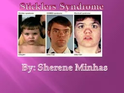
telorism ⢠Type II higher frequency of deafness. ⢠Cochlear deafness â 25 - 70% type i â 50 - 88% type II ⢠Autosomal dominant ⢠(Pax3 mouse gene) on chromosome 2 ⢠http://www.emedicine.com/derm/topic690.htm Larsen ⢠Abnormalities of the growth centres and length of the bones ⢠Short stature ⢠Scoliosis ⢠Short stubby fingers ⢠Broad thumbs ⢠Short metacarpals ⢠Cervical kyphosis ⢠Spina - bifida ⢠Cleft lip and/or palate ⢠Tracheomalacia ⢠Feeding difficulties ⢠Mixed hearing loss ⢠http://www.ncbi.nlm.nih.gov/entrez/disp omim.cgi?id=150250 Cartilage abnormalities with trache
omalacia, cleft palate and hearing loss Ectrodactyly, ectodermal dysplasia and cleft lip/palate ⢠EEC syndrome ⢠Ectrodactyly â Missing or irregular digits ⢠Ectodermal dysplasia ⢠Cleft lip/palate ⢠Chromosome 19, â region defined by D19S894 â http://www.nfed.org/EEC.htm Chondrodysplasia Punctata ⢠Skeletel abnormality ⢠Punctate calcification of the cartilage of the epiphyses, larynx and trachea. ⢠Growth retardation, ⢠Shortening of limbs, ⢠Cataracts, ⢠Dry and scaly skin affects ears ⢠Conradi - hunermann (X - linked dominant form) ⢠http://www.hon.ch/HONselect/RareDiseases/EN/
C05.116.099.708.195.html Osteopetrosis ⢠Recessive form more severe ⢠Sclerotic bones ⢠Long bones may have longitudinal striations ⢠Macrocephaly ⢠Square - shaped head ⢠Frontal bossing ⢠Ptosis ⢠Strabismus ⢠Progressive pressure on cranial nerves leads to â Optic atrophy â Deafness and â Facial palsy ⢠The teeth are abnormal and decay easily ⢠http://www.emedicine.com/med/topic1692.htm Craniometaphyseal dysplasia ⢠Dysplastic disease ⢠Either autosomal dominant or autosomal recessive ⢠Facial deformity ⢠Cranial hyperostosis ⢠Failure of tubular bones to undergo normal modellin
g ⢠Optic nerve atrophy ⢠Compression of the brain stem. ⢠Radiographic features include progressive sclerosis about the cranial sutures and base of the skull and obliteration of the paranasal sinuses . ⢠Facial deformity ⢠Malocclusion of teeth ⢠Nasal obstruction ⢠Facial paralysis ⢠Deafness ⢠Optic nerve atrophy ⢠http://www.medterms.com/script/main/art.asp ?articlekey=17537 Cranial nerve compression and nasal obstruction with facial deformity Achondroplasia ⢠Short stature ⢠Rhizomelic (proximal) shortening of the arms and legs with redundant skin folds on limbs ⢠Limitation of elbow extension
⢠Trident configuration of the hands ⢠Genu varum (bow legs) ⢠Thoracolumbar gibbus in infancy ⢠Exaggerated lumbar lordosis, which develops when walking begins ⢠Large head with frontal bossing ⢠Midface hypoplasia ⢠Obstructive apnea or central apnea â adenotonsillar hypertrophy â hypotonicity â Narrow pns ⢠Frequent otitis media ⢠Sensorineural hearing loss ⢠"towering" petrous ridges â PMID: 8456822 ⢠http://www.emedicine.com/ped/topic12.htm NF type 1 ⢠Autosomal dominant ⢠1 in 3000 individuals. ⢠Cafe au lait patches (more than six greater than 1.5 cms in diameter) â
¢ Peripheral neurofibromata. ⢠Macrocephaly ⢠Short stature ⢠Hypertelorism, and ⢠Thorax abnormalities ⢠Cardiac malformations ⢠Hypertension due to renal artery stenosis ⢠Phaeochromocytoma ⢠Neurofibrosarcomas ⢠Meningiomas ⢠Variable sensorineural deafness ⢠(Acoustic neuromas) probably not ⢠http://www.emedicine.com /ped/topic2418.htm NF type 2 ⢠'Central' form of neurofibromatosis (NF) ⢠The average age of onset is in the early twenties ⢠Presenting symptoms : â vestibular schwannoma (acoustic neuroma âAbâ), â cranial meningioma â spinal tumour. ⢠.ilateral Abâs â¢
Cafe au lait patches may occasionally be present, but are not as numerous as in NF1 ⢠5 - 10% of patients with AN have NF2 ⢠The gene has been mapped to 22q ⢠http://www.emedicine.com/radio/topic475.htm VATER ⢠V: Vertebral dysgenesis ⢠A: Anal atresia ⢠C: Cardiac anomalies ⢠T - E: Fistula +/ - esophageal atresia ⢠R: Renal or Radius anomalies ⢠L: Limb anomalies ⢠S: Single Umbilical artery ⢠http://children.webmd.com/VACTERL - Association McCune - Albright syndrome ⢠At least 2 of the triad â polyostotic fibrous dysplasia â café au lait skin pigmentation â autonomous endocrine hyperfun
ction ⢠Cushing syndrome generally results in growth failure and hypertension in infancy. ⢠The adrenal glands are bilaterally enlarged ⢠Bone changes in the skull can lead to blindness or deafness ⢠http://www.emedicine.com/ped/topic1386.htm Deafness ⢠Patho - emryology ⢠Classification of Childhood Hearing Loss â Congenital without organ involvement â With Associated Organ involvement ⢠Waardenburg ⢠LEOPARD ⢠Skeletal Dysplasia â Klippel - Feil Syndrome â Pierre Robin Syndrome â Van der Hoeve's Syndrome (Osteogenesis imperfecta) ⢠Craniofacial Anomalies â Treacher - Collins Syndrome (Mandi
bulofacial dysostosis) â Goldenhar's Syndrome (Oculoauriculovertebral dysplasia) â Crouzon's Syndrome (Craniofacial dysostosis) â Apert's Syndrome (Acrocephalysyndactyly ⢠Ocular Abnormalities â Usher's Syndrome â Alstrom's Syndrome â Refsum's Disease ⢠Cardiac Abnormalities â Jervell and Lange - Nielsen Syndrome ⢠Renal Abnormalities â Alport's Syndrome ⢠Endocrine Dysfunction â Pendred's Syndrome ⢠Metabolic Storage Diseases ⢠Chromosomal - trisomy21 ⢠Acquired â Prenatal ⢠Maternal ingestion of teratogens/ototoxins ⢠Intrauterine infection â Rubella â CMV â Toxoplasmosis â
Influenza â Herpes 1 and 2 â Syphillis â Postnatal ⢠Prematurity ⢠Anoxia ⢠Kernicterus Pathoembryology ⢠A. Inner ear has separate development (otic placode) at a different time (4th - 20th week gestation) from external and middle ear, however inner ear abnormalities do occur in 15 - 20% of cases of external canal atresia (SNHL in only 6%). ⢠B. Auricle (1st and 2nd branchial arches) is formed early from 6th - 12th week gestation and malformation of auricle implies malformation of middle ear, mastoid, VII nerve. â Normal auricle with external canal (1st branchial groove) atresia indicates abnormal development during 28th wee
k gestation by which time ossicles and middle ear already formed. â Ossicles: 1st arch derivatives -- malleus head, incus short process and body; 2nd arch derivatives -- manubrium of malleus, incus long process, stapes. ⢠C. Histopathology of Congenital SNHL â 1. Abnormal bony labyrinth ⢠a. occurs rarely ⢠b. due to faulty induction of inner ear (bony labyrinth inductor = developing CNS); fetus with abnormal brain development wil l o ften have abnormal bony labyrinth ⢠c. abnormal bony labyrinth seen on radiographic exam of inner ear - - suspect CNS abnormality â 2. Abnormal neuroepithelium ⢠a. occurs more frequently -- predomin
ant histopathology in most congenital and postnatal SNHL ⢠b. usually absence of organ of Corti + abnormal surrounding membranous structures ⢠c. thought to be secondary to premature cell death (degeneration) rather than lack of development ⢠d. with abnormal bony labyrinth, neuroepithelium is often normal ⢠e. with loss of organ of Corti and sound deprivation, normal development of CNS may be impaired; some of the CNS changes (wit h subsequent behavioral deficits) may be ameliorated by early use of sound stimulation -- early detection of hearing loss is imperat ive Genetic deafness ⢠Of all congenital deafness, â 50% is secondary to acquired
disease â 50% is inherited ⢠Of the inherited congenital deafness â 75 - 88% is autosomal recessive (AR), â 12 - 24% is autosomal dominant (AD), â 1% is X - linked ⢠Majority of hereditary congenital HL is clinically undifferentiated (not part of recognizable syndrome and does not affect other organ systems) ⢠Conductive autosomal recessive HL always exists as part of a syndrome; clinically undifferentiated conductive HL is therefore autosomal dominant ⢠Autosomal recessive is usually stable; autosomal dominant tends toward progression Inner Ear Aplasia ⢠1. General â a. inner ear deformity can occur alone or in associati
on with a recognized syndrome â b. more accurately thought of as a spectrum ⢠2. Classification â a. Michel Aplasia (1) most severe form (2) complete failure of bony and membranous labyrinth development (3) no residual hearing â b. Mondini Aplasia ⢠(1) incomplete formation of bony and membranous labyrinth, often asymmetrical ⢠(2) middle and apical turns of cochlea occupy common bony space (cloaca) ⢠(3) cochlea represented by single curved tube with 1 1/2 instead of 2 3/4 turns ⢠(4) vestibular structures also underdeveloped ⢠(5) 2nd most common, (autosomal dominant) â c. Scheibe Aplasia ⢠(1) most common, (autosomal
recessive) ⢠(2) membranous aplasia of pars inferior (cochlea/saccule) ⢠(3) bony labyrinth normal (no radiographic abnormality) ⢠(4) Audio -- profound SNHL with residual low frequency hearing â d. Alexander Aplasia ⢠(1) least severe ⢠(2) partial aplasia of cochlear duct (basal turn) ⢠(3) high frequency HL Associated Organ System Involvement ⢠a. Waardenburg Syndrome â (1) autosomal dominant - variable penetrance â (2) most frequently diagnosed hereditary deafness syndrome (1 - 2% of all congenital deafness) â (3) 6 predominant features: ⢠1. widely spaced medial canthi with shortening of palpebral fissures;
⢠2. prominent, broad nasal route; ⢠3. hypertrichosis of eyebrows; ⢠4. white forelock; ⢠5. heterochromia of irides; ⢠6. SNHL (total or subtotal) in 20% of cases â (4) other features: cleft lip/palate, high arched palate, areas of depigmentation, absent vestibular response â (5) Diagnosis: observing stigmata in patient or family (dystrophic canthi and increased intercanthal distance are most common expressions of syndrome) ⢠b. LEOPARD Syndrome â (1) autosomal dominant â (2) lentigines, EKG abnormality, ocular hypertelorism, pulmonary stenosis, abnormal genitalia, retardation of growth, bilateral sensorineural deafness
(25% affected) Skeletal Dysplasia ⢠a. Klippel - Feil Syndrome â (1) autosomal recessive; females � males â (2) severe progressive SNHL with middle ear anomalies (conductive component), short neck due to fused cervical vertebrae, spina bifida, EAC atresia ⢠b. Pierre Robin Syndrome Sequence â (1) autosomal dominant â (2) main features: cleft palate (50%), micrognathia, glossoptosis â (3) mixed hearing loss, malformed auricles, mental retardation, hypoplastic mandible, aspiration -- common cause of death ⢠c. Van der Hoeve's Syndrome (Osteogenesis imperfecta) â (1) autosomal dominant â (2) bone fragility, blue scler
a, progressive deafness -- conductive, sensorineural, or mixed beginning in childhood (50% affected) Craniofacial Anomalies ⢠a. Treacher - Collins Syndrome (Mandibulofacial dysostosis) â (1) autosomal dominant â (2) auricle malformed and small; preauricular fistulas; EAC atresia â (3) mandibular and malar hypoplasia -- fishmouth â (4) usually bilateral involvement â (5) down sloping palpebral fissures; notched lower lids â (6) CHL due to malformed malleus/incus (stapes normal) -- 30% have significant HL ⢠b. Goldenhar's Syndrome (Oculoauriculovertebral dysplasia) â (1) autosomal dominant or autosomal recessive â (2) 1st
and 2nd arch abnormality â (3) often unilateral (hemifacial microsomia) â (4) malformed auricles/microtia; malformed middle ear/ossicles; possible underdeveloped inner ear â (5) mostly CHL -- 40% affected â (6) upper lid colobomas, epibulbar dermoids, hypoplasia of mandible/ maxilla, cleft palate, spinal anomalies ⢠c. Crouzon's Syndrome (Craniofacial dysostosis) â (1) autosomal dominant â (2) CHL due to EAC atresia or middle ear malformations -- 30% affected â (3) premature closure of cranial suture lines, midface hypoplasia, bulging eyes -- "frog face" ⢠d. Apert's Syndrome (Acrocephalysyndactyly) â (1) autosomal dominant
â (2) facial morphology similar to Crouzon's syndrome, but also syndactyly of hands and feet â (3) mental retardation common Ocular Abnormalities ⢠a. Usher's Syndrome â (1) autosomal recessive â (2) 4% of congenital deafness â (3) characteristics: congenital SNHL, progressive retinitis pigmentosa, night blindness and tunnel vision, cataracts, mental ret ardation, psychosis, spinocerebellar ataxia â (4) vestibular dysfunction -- almost all will have no vestibular response to caloric test or ice water calorics; helps in diagnosi s when etiology unknown â (5) usually born deaf due to atrophy of organ of Corti with progressive vision
loss from retinitis pigmentosa; night blindnes s i s 1st retinal sign (before age 10) with 50% patients having decreased visual acuity by 2nd decade â (6) retinal findings: granular accumulation of pigment beginning at fundus and extending to periphery â (7) Diagnosis: ophthalmoscopy or electroretinography -- perform in infant born with SNHL of unknown etiology; may need routine fun doscopic exams during first 2 decades; important to determine if vision will be lost because if deaf patient loses vision, must initia te special habilitative intervention; important for parents to know if they carry the gene; test family members (audio, electroretinography) to iden
t ify heterozygote carriers ⢠b. Alstrom's Syndrome â (1) autosomal recessive â (2) progressive SNHL beginning in childhood, obesity, diabetes, retinal pigmentation ⢠c. Refsum's Disease â (1) autosomal recessive â (2) progressive aymmetric SNHL (50%), neurologic dysfunction, retinitis pigmentosa, ichthyosis beginning late childhood â (3) absence of enzyme for phytanic acid metabolism (from chlorophyll) ( â 4) Dx.: high phytanic acid plasma level â (5) Rx.: eliminate phytanic acid from diet Cardiac Abnormalities ⢠a. Jervell and Lange - Nielsen Syndrome â (1) autosomal recessive â (2) characteristics: profound
bilateral SNHL (high frequency worst), recurrent syncope (prominent feature), usually terminates in sudden death -- early detection important to preserve life â (3) suspect with family history of SIDS â (4) EKG abnormalities: large T waves, prolonged QT interval â (5) syncopal episodes usually begin 2nd or 3rd decade, last 5 - 10 minutes, frequency variable (EKG -- asystole followed by V. tach) â (6) 50% die before age 15 w/o treatment (7) Treatment: 1. propanolol +/ - phenobarbital 2. implant cardioverter defibrillator when refractory to drugs â (8) Diagnosis: all patients with idiopathic SNHL should have at least 1 EKG; attempt to identify ca
rriers in family members with EKG (note: EKG may normalize in young adults) â (9) cardiac histopathology: hypertrophy of intima of artery to SA node, infarction of SA node, abnormal Purkinje fibers and AV node Renal Abnormalities ⢠a. Alport's Syndrome â (1) autosomal dominant â (2) most common progressive inherited cause of SNHL in children â (3) characteristics: progressive SNHL usually beginning age 10, progressive nephritis with hematuria and proteinuria beginning 1st or 2nd decade â (4) males more severely affected -- if untreated usually die by 3rd decade; females often have normal life span with toxemia during pregnancy â (5)
Diagnosis: consider Alport's in any child with SNHL of recent onset; UA -- �3 RBCs/HPF, �5 WBCs/HPF, proteinuria; serum Cr/BUN abnormal; IVP -- atrophy or lobulated kidney with advanced disease â (6) HL usually progresses with age, predominant in HF, affects 50%, correlation between HL severity and renal disease severity; seldom more than severe HL -- helped significantly with hearing aid; possible improvement in HL after renal dialysis or renal transplant â (7) ocular abnormality may also occur and is important because of possible impairment of 2 sensory modalities (similar to Usher's Syndrome and Congenital Rubella); occurs in 23% with lens
most commonly involved â (8) histopathology: degeneration of organ of Corti, atrophy of spiral ganglion, thickening of glomerular basement membrane, focal sclerosis, interstitial fibrosis, foam cells Endocrine Dysfunction ⢠a. Pendred's Syndrome â (1) autosomal recessive â (2) variable bilateral SNHL (atrophy of organ of Corti) -- usually severe to profound, especially high frequency â (3) develop goiter which usually apparent before age 8; usually euthyroid; no association with cancer; perchlorate test - - abnormal organification of nonorganic iodine â (4) comprise 1 - 7% severe to profoundly deaf children â (5) U - shaped
audiogram, vestibular response variable â (6) Treatment: exogenous thyroid hormone; total or partial thyroidectomy is contraindicated -- goiter returns Metabolic Storage Diseases ⢠a. Mucopolysaccharidoses ⢠(1) ie., Hurler's Syndrome (Gargoylism); Hunter's Syndrome is same but X - linked ⢠(2) characterized by excessive urinary excretion of glycosaminoglycans ⢠(3) abnormal mucopolysaccharides are deposited in tissues ⢠(4) all autosomal recessive (except Hunter's) ⢠(5) hearing loss is conductive or mixed with air - bone gap in high frequencies ⢠(6) dwarfism, forehead prominence, low set ears, mental retardation, hepatosple
nomegaly, coarsening of facial features, corneal opacities ⢠(7) death in early adolescence Chromosomal Abnormalities ⢠a. Trisomy 13 and 18 â lethal in infancy â mixed hearing loss ⢠Trisomy 21 â Mixed hearing loss important to recognize because the sensory deprivation may be significant burden for Down's children) Acquired Congenital Deafness ⢠A. Maternal ingestion of teratogens/ototoxins â 1. Thalidomide â 2. Streptomycin â 3. Quinine, Chloroquine phosphate ⢠B. Intrauterine infection (note: often no signs/symptoms of maternal infection) â 1. Rubella ⢠a. most common cause of acquired congenital SNHL
⢠b. earlier in intrauterine life -- more severe effects, 5 - 10% of mothers with rubella in 1st trimester give birth to children with deafness ⢠c. clinical presentation: (after 1 week of inapparent viral infection) -- posterior cervical and retroauricular adenopathy follo wed by mild URI, then rash which begins on face and disappears rapidly in 72 hrs. ⢠d. Diagnosis: physical exam +immunologic techniques, maternal rash or exposure history ⢠e. Findings: pigmentary retinitis ("salt and pepper") -- most common and consistent finding; congenital cataracts, microcephaly (mental retardation), cardiovascular anomalies, IUGR, jaundice, long bone lesio
ns ⢠f. suggested that all pregnant women have rubella titers drawn at beginning of pregnancy and follow - up titers later ⢠g. Titer: diagnosis based on rubella specific IgM in cord blood or mother/infant serum in 1st 6mos.; older children -- more diffic ult diagnosis (vaccination, infection with wild type virus); titer should be obtained from all patients and mothers when etiology of SNHL unknown (unless child has been immunized) ⢠h. severe to profound SNHL, may be assymetric, may be progressive; may have CHL -- fixed stapes or CSOM secondary to high arched palate, therefore audiometric + impedance monitoring for progressive HL or CHL is important
⢠i. there is an increased incidence of SNHL caused by rubella in families with genetic predisposition to SNHL (possibly predis pos ition increased by presence of both factors) Acquired Congenital Deafness â 2. Cytomegalic inclusion disease ⢠a. CMV -- very ubiquitous viral infection, nearly always without signs/ symptoms of maternal illness;55 - 72% pregnant women have positive CMV titers -- 10% of their offspring have CMV infection; majority of CMV infected infants appear normal but 10 - 15% develop disabilities from CNS damage (developmental delay, learning difficulties); neonates with symptoms at birth (less common) have more severe form and HL is co
mmon (1 in 3000 live births) ⢠b. CMV can infect neonate when mother has a primary or subsequent infection (consecutive pregnancies with infected children); children infected during mother's primary infection have more severe morbidity ⢠c. Teratogenic action variable with lower incidence of pathologic change than rubella -- HL of variable severity, can be progressive ⢠d. Stigmata: microcephaly, hydrocephaly, mental retardation, palatal abnormalities, intracerebral calcifications, prematurity, low birth weight, chorioretinitis, jaundice with hepatosplenomegaly, petechiae ⢠e. Diagnosis: positive titers (CMV specific IgM) from mother and child (somet
imes deceiving), stigmata (may not be present), positive CMV urine cultures (very helpful in early infancy); No established means of prevention or treatment Other Intrauterine Infections ⢠a. Toxoplasmosis -- parasitic disease causing fever, lymphadenopathy, pulmonary lesions and calcific foci in mother ⢠b. Influenza -- maternal fever, lymphadenopathy, URI, arthralgias, myalgias ⢠c. Herpes 1 and 2 -- history of labial or vaginal lesions ⢠d. Syphilis -- 25 - 38% of patients with congenital syphilis have HL â (1) early (infantile) form -- severe, bilateral SNHL, usually multisystem involvement with fatal outcome â (2) late (tardive)
form -- onset in early childhood with bilateral, severe, sudden SNHL associated with vestibular symptoms (similar to Meniere's disease); may have later onset (as late as 5th decade) -- mild HL with severe episodic vertigo; one ear affected first followed by the other ear ( â 3) Histopath: osteitis, obliterative endarteritis, endolymphatic hydrops â (4) Signs: Hennebert's -- positive fistula test with no clinical evidence of middle ear or mastoid disease or fistula (fibrous bands between footplate and vestibular membranous labyrinth or excessively mobile footplate); Tullio's phenomenon -- vertigo and nystagmus with high intensity sound â (5) Treatment: pen
icillin + steroids Perinatal Causes ⢠1. Prematurity â a. most common cause due to factors including: anoxia, kernicterus, ototoxic medication, labyrinthitis, meningitis, temporal bone fractures â b. prematurity as a factor in SNHL -- 10% â c. 2% children less than 1.36 kg (3 lbs.) have significant SNHL â d. premature infant has 20x greater chance of deafness than normal birth weight child â e. usually severe SNHL especially HF f. 68% of premature children with SNHL have multiple handicaps (aphasia, MR, visual pathology, emotional disturbance) and this must be considered in planning rehab program ⢠2. Anoxia â a. most important fa
ctor in SNHL of premature infant; asphyxia likely with cord blood pH .25, 5 minute Apgar â b. pathologic lesion probably lies in cochlear nucleus and other portions of CNS (not cochlea) â c. high rate of other CNS handicaps ⢠3. Kernicterus â a. many are also premature â b. few cases of RH incompatibility, but ABO and other blood group incompatabilties assoc. with kernicterus + SNHL â c. hearing loss -- bilateral HF SNHL with little loss of speech discrimination; 22% have severe SNHL correlating with severity of hemolytic disease â d. Histopath: abnormal CNS with greatest nerve cell injury in ventral cochlear nucleus â e. exchange tr