
24 Department of Neurosurgery Clemens Hospital Academic Hospital of Münster University Münster Germany Department of Radiology Clemens Hospital Academic Hospital of Münster University Müns ID: 944246
Download Pdf The PPT/PDF document "CASE REPORT" is the property of its rightful owner. Permission is granted to download and print the materials on this web site for personal, non-commercial use only, and to display it on your personal computer provided you do not modify the materials and that you retain all copyright notices contained in the materials. By downloading content from our website, you accept the terms of this agreement.


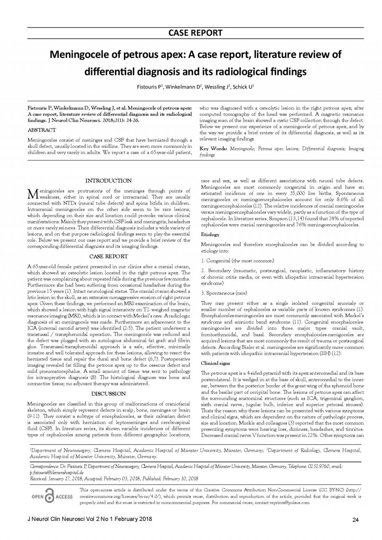
CASE REPORT 24 Department of Neurosurgery, Clemens Hospital, Academic Hospital of Münster University, Münster, Germany; Department of Radiology, Clemens Hospital, Academic Hospital of Münster University, Münster, Germany.Correspondence: Dr. Fistouris P, Department of Neurosurgery, Clemens Hospital, Academic Hospital of Münster University, Münster, Germany, Telephone: 0251.976-0, email: p.fistouris@clemenshospital.de This open-access article is distributed under the terms of the Creative Commons Attribution Non-Commercial License (CC BY-NC) (http://creativecommons.org/licenses/by-nc/4.0/), which permits reuse, distribution and reproduction of the article, provided that the original work is properly cited and the reuse is restricted to noncommercial purposes. For commercial reuse, contact reprints@pulsus.com Meningocele of petrous apex: A case report, literature review of di�eren�al diagnosis and its radiological �ndingsFistouris P, Winkelmann D, Wessling J INTRODUCTIONeningoceles are protrusions of the meninges through points of weakness, either in spinal cord or intracranial. They are usually connected with NTDs (neural tube defects) and spina bifida in children. Intracranial meningoceles on the other side seem to be rare lesions, which depending on their size and location could provoke various clinical manifestations. Mainly they present with CSF leak and meningitis, headaches or more rarely seizures. Their differential diagnosis includes a wide variety of lesions, and on that purpose radiological findings seem to play the essential role. Below we present our case report and we provide a brief review of the CASE REPORTA 65-year-old female patient presented in our clinics after a cranial ct-scan, which showed an osteolytic lesion located in the right petrous apex. The J Neurol Clin Neurosci Vol 2 No 1 February 2018 be sixth cranial nerve palsy and diplopia. Symptoms often present months or Diagnosis and management of petrous apex lesion present unique challenges secondary to their centralized location and their anatomical adjacements. Imaging studies (CT-Scan, MRI) are the primary method used to diagnose these lesions. Subsequently we report a differential diagnosis of petrous apex 3. Neoplasms (such as Chordosarcoma, Chordoma, meningeoma, metastasis)More specially and regarding cephaloceles-meningoceles we have to take into consideration the non-neoplasmatic lesions. Thy are representing an amount of more than 90% of all pe
trous apex lesions. Depending on pathophysiology, the majority of nonneoplastic petrous apex lesions are inflammatory complications of air cell diesease. Pneumatization of petrous apex occurs when epithelium-lined air cells develop as medial communication from the mastoid air cells. The air cells of the petrous apex are susceptible to similar pathologic processes that occur in the mastoid segment including A low-dose non-contrast CT scan (80 mA & 120 kV) was performed with a Toshiba Aquilion CX CT helical scanner with wide window width (2700 HU) and level (400 HU) using a bone filter kernel. MR imaging was performed on a Siemens Skyra 3-Tesla MR unit with standard T1-weighted (spin echo; TR 600 ms/TE 20 ms; 5 mm slice thickness), T2-weigted 3d (spin echo; TR 1400 ms/ TE 152 ms; 0,8 mm slice thickness) and T2-weighted inversion recovery (spin-echo; TR 9000 ms/ TE 81 ms; 5 mm slice thickness) sequences; additional diffusion weighted imaging (RESOLVE DWI; TR 6070 ms/ TE 65 ms, b=0 & 1000; 4 mm slice thickness), and a contrast-enhanced T1 weighted 3d (gradient echo [MP RAGE]; TR 2200 ms/ TE 2,45 ms; 1 mm slice thickness) sequence was executed. The CT scan shows a unilateral expansile cyst with a maximum diameter of 21 mm and thinned sclerotic but sharply defined osseous margins in the right petrous apex, the body of the sphenoid bone and the clivus (Figure 1). The lesion erodes the bone to the posterior portion of Meckel’s cave, the carotid canal wall and the petrooccipital fissure. The cyst content has got a homogenous fluid-like attenuation (10-20 HU). T1-weighted MR images show a homogenous low signal, non-enhancing cyst content and a thin wall of low signal intensity encircling the cyst, with mild rim enhancement. T2-weighted MR images reveal fluid-like homogeneous hyperintensity. There is no diffusion restriction within the cyst. The cyst neither extends into the cerebellopontine angle nor into the inner or middle ear structures. In the contrast enhanced MR images and the 3d T2-weighted images the cyst appears related with Axial bone algorithm CT scan shows a unilateral expansile cyst extending Figure 2a) Axial non-contrast T1-weighted SE (TR 600ms/ TE 10ms/ slice thickness: 5 mm) MR image reveals a homogenous low signal process, like a cyst (arrow) Petrous apex lesionInflammatory/CysticCharacteristics/PathophysiologyCholesterin granulom/XanthogranulomCholesterin crystal, Foam cells. Anaerobiccatabolism of blood and blood products.MucoceleCholosteatoma/Epid
ermoidObstructed mucosalized air space.Epithelial cell remnants/multipotential embryoniccells.Effusion or retained�Infection of middle ear or mastoid - trapping ofresidual fluid in the petrous apex.Cerebrospinal fluid cysts and cephalocelesMeningocele/CSF cyst/arachnoid cystPoints of weakness, splitting of meninges, skullbase.Cephaloceles/Meckel’s cave diverticulumProtrusion of arachnoid or dura from Meckel’scave.Abszess/petrous apicisSpread of infection through preformed air-cell tractsin temporal bone/usual otitis media.AneurysmaPetrous carotis interna aneurysma/posttraumatic,congenital, mycotic.Asymmetric pneumatizationAsymmetrie of air cells (5-10 % of all individualls).10-30 % air cells in temporal bones.TABLE 1 Differential diagnosis of these lesions J Neurol Clin Neurosci Vol 2 No 1 February 2018 26 Naidich TP, Altman NR, Braffman BH, et al. Cephaloceles and re- lated Moore KR, Harnsberger HR, Shelton C, et al. ‘Leave me alone’ le- sions Muckle RP, Cruz A, Lo WM. Petrous apex lesions. Am J Otol. Isaacson B, Kutz JW, Roland PS. Lesions of Petrous Apex : Diagnosis and Kevin RM, Nancy JF, Harnsberger HR, et al. Petrous Apex Cephaloceles. Çavusoglu M, Duran S, Hatipoglu HG, et al. Petrous apex cephalocoele: contribution of coexisting intracranial pathologies to the Perez JA, Juncos JMM. Parietal intradiploic encephalocele: Report of a Alorainy IA. Petrous apex cephalocele and empty sella: Is there any Chapman PR, Shah R, Curé JK, et al. Petrous Apex Lesions: Pictorial Kou YF, Allen KP, Isaacson B. Recurrent meningitis secondary to a 11.Cohen MM, Lemire RJ. Syndromes with cephaloceles. Teratology. Bialer OY, Rueda MP, Bruce BB, et al. Meningoceles in idiopathic Jimenez DF, Barone CM. Encephaloceles, Meningoceles and Dermal Simpson DA, David OJ, White J. Cephaloceles: treatment, outcome and Figure 2c) Axial T2-weighted 3d SE (TR 1400 ms/ TE 152ms/ slice thickness: 0,8 mm) MR image reveals a cyst (asterisk), that appears related with Meckel’s cave(arrow) Axial contrast-enhanced fatsaturated T1-weighted GE (TR 2200ms/ TE 2,45ms; slice thickness: 1 mm) MR image shows a low-signal-intensity cyst with mild Figure 2b) Axial DWI [diffusion weighted imaging] (TR 6070ms/ TE 65ms/ slice thickness: 4 mm) MR image demonstrates no diffusion restriction within the lesion J Neurol Clin Neurosci Vol 2 No 1 February 2018 Meningocele of petrous apex: A case report, literature review of differential diagnosis and its radiological �nding