
and Blood V essels Crash Course httpswwwyoutubecomwatchv9fxm85Fy4sQampindex27amplistPL3EED4C1D684D3ADF The circulatory system is the system that transports materials around the body to and from the cells ID: 998510
Download Presentation The PPT/PDF document "Circulatory System H eart" is the property of its rightful owner. Permission is granted to download and print the materials on this web site for personal, non-commercial use only, and to display it on your personal computer provided you do not modify the materials and that you retain all copyright notices contained in the materials. By downloading content from our website, you accept the terms of this agreement.


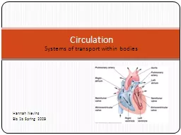
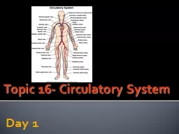

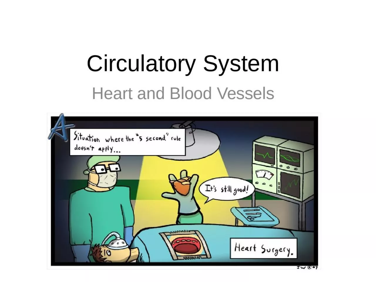
1. Circulatory SystemHeart and Blood Vessels
2. Crash Coursehttps://www.youtube.com/watch?v=9fxm85Fy4sQ&index=27&list=PL3EED4C1D684D3ADF
3. The circulatory system is the system that transports materials around the body to and from the cells.
4. Question: Why do humans need a circulatory system whereas bacteria and simple organisms do not?Answer: Because a complex organism such as a human has many cells that are far from the outside environment where nutrients would come from. The system brings the materials to the cells that would not normally receive them.
5. Humans have a closed circulatory system: This means that the blood is always contained in tubes and vessels.
6.
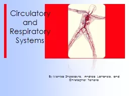
7. Parts of the Circulatory SystemThe human circulatory system is composed of the following: 1. Blood Vessels 2. Heart 3. Blood
8. Blood VesselsArteriesStructure:Thick, elasticSmallest arteries are called arteriolesDO NOT CONTAIN VALVESFunction: Transport blood AWAY from the heart to the organs and tissues of the body.
9. Blood VesselsVeinsStructure:Thin and slightly elasticSmallest veins are called venulesContain VALVES – to help blood flow back to the heart against the force of gravity. Lower pressure than arteriesFunction: To RETURN blood from the body tissues to the heart.
10. Blood VesselsCapillariesStructure: Microscopic blood vessels that connect arterioles and venulesThin walled and narrowBlood cells pass through them in single file Function: Allows material and gas exchange between the body cells and the blood.
11.
12. The HeartStructure: A four chambered muscular organ located in the chest cavity of a human. Made of cardiac muscle. It is covered by a pericardium that protects it. Pericardium: A tough membrane that surrounds the heart.
13.
14. Function: Pump blood around the body supplying the cells with nutrients and removing wastes (CO2) from the cells.
15.
16.
17.
18.
19.
20.
21.
22.
23.
24.
25.
26.
27.
28.
29.
30. Video Time http://studyjams.scholastic.com/studyjams/jams/science/human-body/circulatory-system.htm
31. Blood Flow through the HeartDeoxygenated blood from the body enters the ______________via the __________and ______________________. Here the blood is passed through the _____________to the _______________.right atriuminferiorsuperior vena cavatricuspid valveright ventricle
32. The right ventricle contracts and forces blood up through the ___________ valves and out through the left and right _________ arteries.This brings blood to the ________ to be oxygenated. semilunarpulmonarylungs
33. Oxygenated blood from the lungs returns to the heart via the left and right ___________ veins to the ____________.The blood is passed to the _____________ through the __________ valve.The left ventricle contracts and pushes blood through the semilunar valves and out through the _______ to the body. pulmonaryleft atriumleft ventriclebicuspidaorta
34. The Heartbeat CycleA single beat of the heart consists of contractions of the atria followed by contractions on the ventricles and then a period of relaxation of all four chambers of the heart.Contractions are called systoles, and relaxations are called diastoles.
35.
36. Video Timehttps://www.youtube.com/watch?v=jLTdgrhpDCg
37. The “LubDub” sound of the HeartbeatThe “LubDub” sound of the heartbeat is caused by the closing of the heart’s valves. Lub Sound -- caused by the closing of the A-V valves (tricuspid, bicuspid). Dub Sound -- caused by the closing of the semilunar valves.
38. Heart Soundshttp://depts.washington.edu/physdx/heart/demo.html
39. Crash Coursehttps://www.youtube.com/watch?v=X9ZZ6tcxArI
40. CONTROL OF THE HEARTBEATThe heart is caused to beat regularly by a structure called the _______________ (S - A node) or the _____________. sinoatrial nodePACEMAKER
41. How it HappensAn electrical impulse from the brain is received by the S-A node (pacemaker) in the right atrium. The impulse spreads quickly over both atria causing atria muscles to contract. The impulse reaches the A-V node (atrioventricular node) in the right ventricle.
42. The pacemaker controls the heartbeat for a human for their entire life. The impulse is transmitted by the AV node down a nerve bundle (bundle of His). The impulse branches to both ventricles causing contraction
43. Q. What happens if the pacemaker gives out? A. The person’s heart will stop beating because the atria and ventricles are not receiving electrical impulses causing them to contract.
44. A person whose pacemaker gives out can get an ___________ one inserted into their chest. artificial
45. CONTROL OF THE HEART RATEThe heart rate (speed) at which the heart beats is controlled by two nerves. ______________________ in the Medulla Oblongata: Nerve in the brain that causes the heart to speed up when needed. ____________: Nerve in the brain that causes the heart to slow down when neededCardioaccelerator nerveVagus nerve
46.
47. The medulla sends a message to the SA node to cause an impulse to be sent to the AV node causing the heart to contract more or less in an attempt to set the heart rate.
48. BLOOD PRESSUREBlood Pressure: A measure of the pressure blood exerts on the walls of blood vessels.
49. Q. How is blood pressure measured? A. Blood pressure is measured using a blood pressure cuff or Sphygmomanometer.
50. It measures the pressure in an artery at its highest point after contraction (systolic pressure) and the lowest point while the heart is resting (diastolic pressure). A simple fraction is calculated using the following formula: Blood Pressure =
51. For example: A person with a pressure 120/80 means that the person has a pressure of 120 while the heart is contracting and 80 when the heart is relaxing. P.S. Normal blood pressure is different for each person but is usually around 120/80. (high is over 140/90)
52. Divisions of Circulation
53. There are two types of circulation that happen in the human organism. Pulmonary circulation Systemic circulation
54. Pulmonary Circulation
55. Systemic Circulation
56. Coronary Circulation
57. Hepatic-portal Circulation
58. Renal Circulation
59.
60. Coronary Artery Supplies blood to nourish the heart muscles
61. Coronary Sulcus Encircles the heart like a crown (corona = crown). The atria are found above and the ventricles are found below this blood vessel.
62. What happens when there is a blockage within the coronary artery?The blood flow to the heart muscles will be blocked, leading to a heart attack.
63. Anterior Interventricular Sulcus Marks the front (ventral) position of the heart. This vessel follows the septum that separates the right and left ventricles.
64. ApexThe pointed end of the heart. Only the left ventricle forms the heart apex.
65. PericardiumThe double-walled sac around the heart. Function: Protects and anchors the heartPrevents overfilling of the heart with bloodAllows for the heart to work in a relatively friction-free environment
66.
67. MyocardiumCardiac muscle layer forming the bulk of the heart
68. Why do you think there is a thicker layer of myocardium in the ventricles than compared to the atria?
69. Why do you think there is a thicker layer of myocardium in the left ventricle than compared to the right ventricle?