
stimulation Enhances Myeloid Cell Activation and Anti Tumor Immunity in the Setting of PD L1 and TIGIT Checkpoint Blockade Abstract Background Co inhibition of TIGIT and PD 1 L 1 impro ID: 940688
Download Pdf The PPT/PDF document "LIGHT TNFSF14 Co" is the property of its rightful owner. Permission is granted to download and print the materials on this web site for personal, non-commercial use only, and to display it on your personal computer provided you do not modify the materials and that you retain all copyright notices contained in the materials. By downloading content from our website, you accept the terms of this agreement.


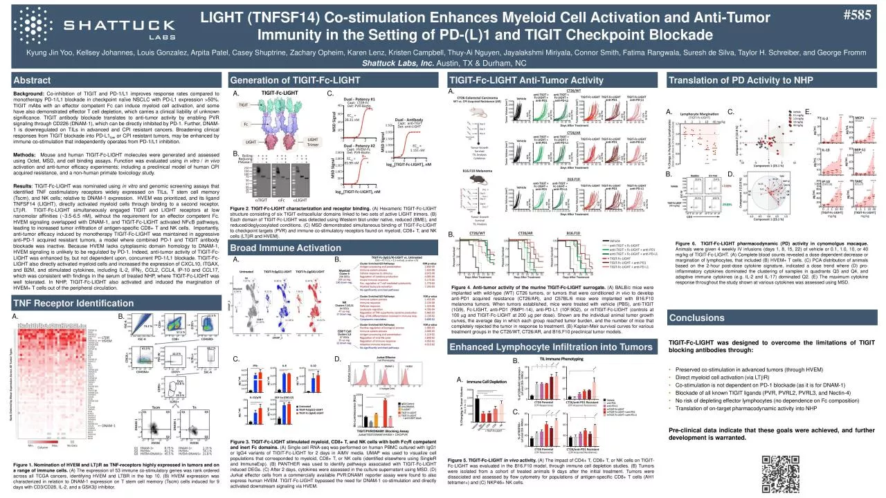
LIGHT (TNFSF14) Co - stimulation Enhances Myeloid Cell Activation and Anti - Tumor Immunity in the Setting of PD - (L)1 and TIGIT Checkpoint Blockade Abstract Background : Co - inhibition of TIGIT and PD - 1 /L 1 improves response rates compared to monotherapy PD - 1 /L 1 blockade in checkpoint naïve NSCLC with PD - L 1 expression � 50 % . TIGIT mAbs with an effector competent Fc can induce myeloid cell activation, and some have also demonstrated effector T cell depletion, which carries a clinical liability of unknown significance . TIGIT antibody blockade translates to anti - tumor activity by enabling PVR signaling through CD 226 (DNAM - 1 ), which can be directly inhibited by PD - 1 . Further, DNAM - 1 is downregulated on TILs in advanced and CPI resistant cancers . Broadening clinical responses from TIGIT blockade into PD - L 1 low or CPI resistant tumors, may be enhanced by immune co - stimulation that independently operates from PD - 1 /L 1 inhibition . Methods : Mouse and human TIGIT - Fc - LIGHT molecules were generated and assessed using Octet, MSD, and cell binding assays . Function was evaluated using in vitro / in vivo activation and anti - tumor efficacy experiments ; including a preclinical model of human CPI acquired resistance, and a non - human primate toxicology study . Results : TIGIT - Fc - LIGHT was nominated using in vitro and genomic screening assays that identified TNF costimulatory receptors widely expressed on TILs, T stem cell memory ( Tscm ), and NK cells ; relative to DNAM - 1 expression . HVEM was prioritized, and its ligand TNFSF 14 (LIGHT), directly activated myeloid cells through binding to a second receptor, LT b R . TIGIT - Fc - LIGHT simultaneously engaged TIGIT and LIGHT receptors at low nanomolar affinities (~ 3 . 5 - 6 . 5 nM ), without the requirement for an effector competent Fc . HVEM signaling overlapped with DNAM - 1 , and TIGIT - Fc - LIGHT activated NF k B pathways, leading to increased tumor infiltration of antigen - specific CD 8 + T and NK cells . Importantly, anti - tumor efficacy induced by monotherapy TIGIT - Fc - LIGHT was maintained in aggressive anti - PD - 1 acquired resistant tumors, a model where combined PD - 1 and TIGIT antibody blockade was inactive . Because HVEM lacks cytoplasmic domain homology to DNAM - 1 , HVEM signaling is unlikely to be regulated by PD - 1 . Indeed, anti - tumor activity of TIGIT - Fc - LIGHT was enhanced by, but not dependent upon, concurrent PD - 1 /L 1 blockade . TIGIT - Fc - LIGHT also directly activated myeloid cells and increased the expression of CXCL 10 , ITGAX, and B 2 M, and stimulated cytokines, including IL - 2 , IFN g , CCL 2 , CCL 4 , IP - 10 and CCL 17 , which was consistent with findings in the serum of treated NHP, where TIGIT - Fc - LIGHT was well tolerated . In NHP, TIGIT - Fc - LIGHT also activated and induced the margination of HVEM+ T cells out of the peripheral circulation . Figure 2 . TIGIT - Fc - LIGHT characterization and receptor binding . (A) Hexameric TIGIT - Fc - LIGHT structure consisting of six TIGIT extracellular domains linked to two sets of active LIGHT trimers . (B) Each domain of TIGIT - Fc - LIGHT was detected using Western blot under native, reduced (BME), and reduced/ deglycosylated conditions . (C) MSD demonstrated simultaneous binding of TIGIT - Fc - LIGHT to checkpoint targets (PVR) and immune co - stimulatory receptors found on myeloid, CD 8 + T, and NK cells ( LT b R and HVEM) . Figure 5 . TIGIT - Fc - LIGHT in vivo activity . (A) The impact of CD 4 + T, CD 8 + T, or NK cells on TIGIT - Fc - LIGHT was evaluated in the B 16 . F 10 model, through immune cell depletion studies . (B) Tumors were isolated from a cohort of treated animals 9 days after the initial treatment . Tumors were dissociated and assessed by flow cytometry for populations of antigen - specific CD 8 + T cells (AH 1 tetramer+) and (C) NKP 46 + NK cells . ⢠Preserved co - stimulation in advanced tumors (through HVEM) ⢠Direct myeloid cell activation (via LT b R ) ⢠Co - stimulation is not dependent on PD - 1 blockade (as it is for DNAM - 1 ) ⢠Blockade of all known TIGIT ligands (PVR, PVRL 2 , PVRL 3 , and Nectin - 4 ) ⢠No risk of depleting effector lymphocytes (no dependence on Fc composition) ⢠Translation of on - target pharmacodynamic activity into NHP Kyung Jin Yoo , Kellsey Johannes, Louis Gonzalez, Arpita Patel, Casey Shuptrine , Zachary Opheim , Karen Lenz, Kristen Campbell, Thuy - Ai Nguyen, Jayalakshmi Miriyala , Connor Smith, Fatima Rangwala , Suresh de Silva, Taylor H. Schreiber, and George Fromm Figure 4 . Anti - tumor activity of the murine TIGIT - Fc - LIGHT surrogate . (A) BALB/c mice were implanted with wild - type (WT) CT 26 tumors, or tumors that were conditioned in vivo to develop anti - PD 1 acquired resistance (CT 26 /AR), and C 57 BL/ 6 mice were implanted with B 16 . F 10 melanoma tumors . When tumors established, mice were treated with vehicle (PBS), anti - TIGIT ( 1 G 9 ), Fc - LIGHT, anti - PD 1 (RMP 1 - 14 ), anti - PD - L 1 ( 10 F . 9 G 2 ), or mTIGIT - Fc - LIGHT (controls at 100 m g and TIGIT - Fc - LIGHT at 200 m g per dose) . Shown are the individual animal tumor growth curves, the average day in which each group reached tumor burden, and the number of mice that completely rejected the tumor in response to treatment . (B) Kaplan - Meir survival curves for various treatment groups in the CT 26 /WT, CT 26 /AR, and B 16 . F 10 preclinical tumor models . Figure 1 . Nomination of HVEM and LT b R as TNF - receptors highly expressed in tumors and on a range of immune cells . (A) The expression of 53 immune co - stimulatory genes was rank ordered across all TCGA cancers, identifying HVEM and LTBR in the top 10 . (B) HVEM expression was characterized in relation to DNAM - 1 expression on T stem cell memory ( Tscm ) cells induced for 9 days with CD 3 /CD 28 , IL - 2 , and a GSK 3 b inhibitor . #585 Shattuck Labs, Inc. Austin, TX & Durham, NC Generation of TIGIT - Fc - LIGHT Translation of PD Activity to NHP TIGIT - Fc - LIGHT Anti - Tumor Activity TNF Receptor Identification Conclusions TIGIT - Fc - LIGHT was designed to overcome the limitations of TIGIT blocking antibodies through : Pre - clinical data indicate that these goals were achieved, and further development is warranted . A. B. A. B. C. Broad Immune Activation Figure 3 . TIGIT - Fc - LIGHT stimulated myeloid, CD 8 + T, and NK cells with both Fc g R competent and inert Fc domains . (A) Single cell RNA - seq was performed on human PBMC cultured with IgG 1 or IgG 4 variants of TIGIT - Fc - LIGHT for 2 days in AIMV media . UMAP was used to visualize cell populations that corresponded to myeloid, CD 8 + T, or NK cells (identified elsewhere using SingleR and ImmuneExp ) . (B) PANTHER was used to identify pathways associated with TIGIT - Fc - LIGHT induced DEGs . (C) After 2 days, cytokines were assessed in the culture supernatant using MSD . (D) Jurkat effector cells from a commercially available PVR : DNAM 1 reporter assay were found to also express human HVEM . TIGIT - Fc - LIGHT bypassed the need for DNAM - 1 co - stimulation and directly activated downstream signaling via HVEM . A. C. B. D. Figure 6 . TIGIT - Fc - LIGHT pharmacodynamic (PD) activity in cynomolgus macaque . Animals were given 4 weekly IV infusions (days 1 , 8 , 15 , 22 ) of vehicle or 0 . 1 , 1 . 0 , 10 , or 40 mg/kg of TIGIT - Fc - LIGHT . (A) Complete blood counts revealed a dose dependent decrease or margination of lymphocytes, that included (B) HVEM+ T cells . (C) PCA distribution of animals based on the 2 - hour post - dose cytokine signature, indicated a dose trend where (D) pro - inflammatory cytokines dominated the clustering of samples in quadrants Q 3 and Q 4 , and adaptive immune cytokines ( e . g . IL - 2 and IL - 17 ) dominated Q 2 . (E) The maximum cytokine response throughout the study shown at various cytokines was assessed using MSD . A. B. C. D. E. Enhanced Lymphocyte Infiltration into Tumors A. B. A. B. C.