
secretory products into the oral cavity the salivary glands In the lab you will also have the opportunity the examine one other specialized epithelial area the lip The oesophagus is the first part of the alimentary canal Its ID: 918343
Download Presentation The PPT/PDF document "ORAL CAVITY The oral cavity is formed by..." is the property of its rightful owner. Permission is granted to download and print the materials on this web site for personal, non-commercial use only, and to display it on your personal computer provided you do not modify the materials and that you retain all copyright notices contained in the materials. By downloading content from our website, you accept the terms of this agreement.


Slide1
Slide2ORAL CAVITY
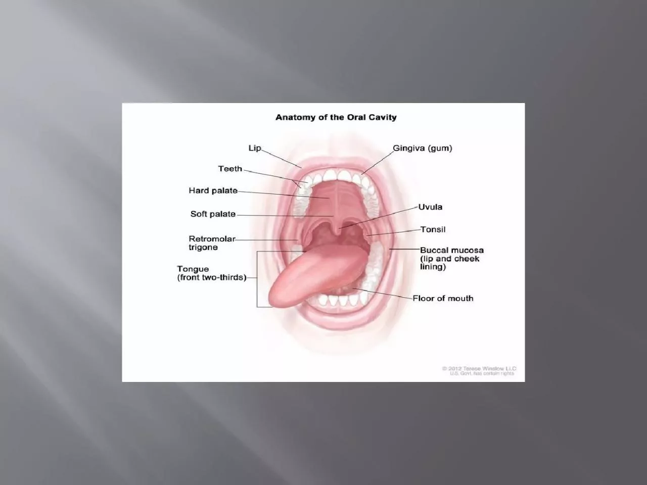
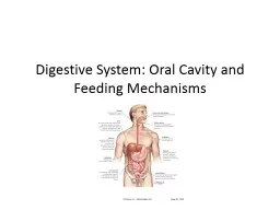
The oral cavity is formed by a bewildering array of tissues which function in or are associated with the processes that are performed with what we typically refer to as our mouth. Lecture and lab focus on the one organ found within the oral cavity, the tongue, and the glands which empty their
secretory
products into the oral cavity, the salivary glands. In the lab you will also have the opportunity the examine one other specialized epithelial area, the lip. The
oesophagus
is the first part of the alimentary canal. Its
organisation
is also typical for all parts of the gastrointestinal tract (GIT).
The oral cavity is divided in a vestibule, the area "outside" the teeth, and an oral cavity proper. The entire oral cavity is lined by a stratified
squamous
epithelium. The epithelial lining is divided into two broad types:
Slide3Masticatory
epithelium covers the surfaces involved in the processing of food (tongue,
gingivae
and hard palate). The epithelium is keratinized to different degrees depending on the extent of physical forces exerted on it.
2-Lining epithelium, i.e. non-
keratinised
stratified
squamous
epithelium, covers the remaining surfaces of the oral cavity
Slide4Tongue
The dorsal surface of the tongue is divided by the
sulcus
terminalis into an oral part, the anterior two-thirds, and a pharyngeal part, the posterior one-third. The dorsal surface of the oral part has a characteristic appearance due to the presence of a large number of small projections, the lingual papillae. The epithelium of the pharyngeal part forms a somewhat irregular surface which covers the lingual tonsils.
Slide5Filiform
papillae
are the smallest and most numerous papillae. By providing the tongue with a rough surface they aid in the manipulation and processing of foods.
Prof. Oxnard brought another function to my attention, i.e.
The lingual papillae consist of a connective tissue core covered with a stratified squamous
epithelium. On the basis of their appearance four types of papillae can be distinguished -
filiform
,
fungiform
,
circumvallate
and foliate papillae.
the cleaning of the surfaces of the mouth, in particular the teeth.
Slide6Fungiform
papillae
occur singly and are fairly evenly spaced between the
filiform
papillae. Their connective tissue core is richly
vascularised
. The epithelium is slightly thinner than on the remaining surface of the tongue.
Circumvallate
papillae
are the largest and least numerous papillae - in humans there are between 8 and 12 of them. They occur in depressions of the surface of the tongue and are surrounded with a trench formed by the
infolding
of the epithelium. Taste buds are particularly numerous on the lateral surfaces of these papillae. The excretory ducts of serous glands open into the trenches surrounding the papillae ("rinsing glands" or glands of von
Ebner
).
Slide7F
oliate papilla
are not well developed in humans and may be absent in aged individuals. If present, they form lamellae along the posterior and lateral border of the tongue.
The epithelium of the dorsal surface of the tongue rests on a fairly dense layer of connective tissue, which connects the epithelium firmly with the underlying muscular and connective tissues
.
Slide8The muscles of the tongue (skeletal muscle) are organized into strands oriented more or less perpendicular to each other. Their actions provide the tongue with the necessary motility to participate in the formation of speech and to aid in the initial processing of foods. Control of the movement of the tongue muscles and the collection of sensory information necessitate a profuse
innervation
of the tongue in which a number of the cranial nerves participate (V, trigeminal nerve - sensory - anterior two-thirds; VII, facial nerve - taste; IX,
glossopharyngeal
nerve - sensory/taste - posterior one-third; XII, hypoglossal nerve - motor).
Slide9Taste Buds
Taste buds are most numerous in the
fungiform
,
circumvallate and foliate papillae. In addition, taste buds are found in the palate, palatoglossal and
palatopharyngeal
arches and in the pharynx and larynx.
In histological sections they appear as ovoid lightly stained bodies, which extend perpendicular from the basement membrane to a little opening formed in the epithelium, the taste pore. The elongated cells that form the taste bud can functionally be divided into three groups: sensory cells, supporting (or
sustentacular
) cells, and basal cells. Sensory cells extend
microvilli
into the taste pore. These
microvilli
contain the receptors for the different basic taste modalities (sweet, salty, bitter and acid). Basal cells regenerate the two other cell types
Slide10Slide11Salivary Glands
Saliva is a mixed secretion, which is derived from numerous large and small salivary glands that all open into the oral cavity. Small salivary glands are situated in the connective tissue beneath the epithelia lining the oral cavity, and, in the case of the tongue, they may also be found between the muscular tissue. Depending on the
localisation
they are grouped into lingual, labial,
buccal, molar and palatine glands.
The large salivary glands form three paired groups:
the sublingual glands, which are positioned beneath the tongue and embedded deeply in the connective tissue of the oral cavity,
the
submandibular
glands and
the parotid glands, which lie outside the oral cavity.
Slide12All of these glands are
tubuloacinar
glands, i.e. they have
secretory
acini but the first part of the duct system originating from the acini also participates in the secretory
process. The salivary glands are divided by connective tissue septa into lobes, which are further subdivided into lobules.
Functionally the
secretory
acini
can be divided into two groups: those that secrete a rather liquid product - serous
acini
, and those that secrete a very viscous product - mucous
acini
. This functional differentiation is reflected in the appearance of these
acini
in histological sections.
Slide13Slide14The cells forming the serous
acini
contain a round or slightly ovoid nucleus which is placed basally in the cell. In an H&E stain, the apical cytoplasm may appear pinkish/red or, in well-preserved tissue, contain reddish granules. The granules represent the vesicles which contain the
secretory
products of the cell. The digestive enzyme α-amylase is also secreted by the acinar cells.
The cells forming the mucous
acini
usually contain flattened nuclei which appear to be "pressed" against the basal surface of the cell.
Secretory
vesicles fill much of the apical cytoplasm. The
secretory
product has either been dissolved during the staining process or remains unstained. Small amounts of cytoplasm which remain between the vesicles gives the apical part of the cell a distinct "spongy" appearance.
Slide15Occasionally, and in particular in glands located relatively close to the oral cavity, serous cells and mucous cells may form compound or mixed
acini
. The serous cells form in these cases small half-moon or crescent-shaped structures, which attach to mucus producing
acini
and empty their secretory product into interstices between the mucus-producing cell. Following their appearance they are called serous
demilunes
.
Slide16Both serous and mucous
acini
and parts of the
secretory
duct system are surrounded by myoepithelial cells which by their contraction participate in the secretory process. They are usually difficult to distinguish in histological sectionsGlands located close to the oral cavity have mainly mucous secretions, whereas glands located further away from the oral cavity have mainly serous secretions. Following this general rule, the parotid glands contain almost exclusively serous acini, the submandibular glands contain both serous and mucous acini
, and the sublingual glands contain mainly mucous acini
or mucous
acini
with serous
demilunes
.
Slide17Slide18The teeth are classified as incisors, canines, premolars, and molars. The eight incisors cut food by their edges. The four canines ("
cuspids
" or "eye-teeth") assist in cutting. The eight premolars ("bicuspids") assist in crushing food. They replace the deciduous molars. The twelve molars crush and grind food. The roots of the upper molars are closely related to the floor of the maxillary sinus. Hence
pulpal
infection may cause sinusitis, or sinusitis may cause toothache. The third molar ("wisdom tooth") is highly variable.
Slide19Primary or deciduous dentition
No functioning teeth have penetrated the oral cavity (i.e., erupted) at birth. The "milk teeth" appear in the oral cavity between the ages of 6 months and 2.5 years. The first teeth to erupt are the lower medial incisors, at about 6 months. All of the deciduous teeth have been shed by about 12 years.
Permanent dentition
The so-called permanent teeth begin to appear in the oral cavity at the age of about 6 years, and they have replaced the deciduous teeth by about 12 years. The first to erupt is the first molar, at about 6 to 7 years (the 6-year molar); the second molar erupts at about age 12 (the 12-year molar); the third molar ("wisdom tooth") may erupt from 17 years onward, or not at all. The 32 permanent teeth are arranged in quadrants of 8 each: 2 incisors, 1 canine, 2 premolars, and 3 molars. The permanent molars have no deciduous predecessors
Slide20. A, Longitudinal section of an incisor. B, Cross-section of the crown of an incisor, showing enamel, dentin, and pulp.
C, Side view
of an incisor, showing the area of epithelial attachment and the line of the
cemento
-enamel junction. D, Longitudinal section of a molar, showing the bifurcation of the pulp cavity