
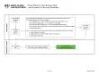
Page 1 of 3 and Aspirate Collection Guideline Preparation Peripheral Smear Clearly identify patient and procedure Assemble collection materials Make 2 direct smears manually adjusting as necessa ID: 936682
Download Pdf The PPT/PDF document "Bone Marrow Core Biopsy Clot" is the property of its rightful owner. Permission is granted to download and print the materials on this web site for personal, non-commercial use only, and to display it on your personal computer provided you do not modify the materials and that you retain all copyright notices contained in the materials. By downloading content from our website, you accept the terms of this agreement.


Page 1 of 3 Bone Marrow Core Biopsy, Clot, and Aspirate Collection Guideline Preparation Peripheral Smear Clearly identify patient and procedure Assemble collection materials Make 2 direct smears manually, adjusting as necessary for proper length and thickness. Perform nger stick Proceed to page 2, Bone Marrow Aspirate Slides Use syringes not rinsed with heparin for slide preparation and clot. Assemble collection tubes. The standard bone marrow collection consists of: Empty tube with cap: ½ mL for clot (drawn in blank syringe with no heparin in it) • One lavender top (EDTA) tube: 3 mL for possible molecular testing • One yellow top (ACD solution B) tube: 4 mL for possible ow cytometric testing • One green top (sodium heparin) tube: 3 mL for possible chromosome analysis and/or FISH testing • Two formalin containers Place 10 clean slides on the work surface for collection. Have other slides available for use if needed. • 2 slides (peripheral blood smear) • 5 slides (bone marrow aspirate: 2 direct preps and 3 unit preps) • 3 slides (biopsy touch preps) Label slides, tubes, and containers MC4091-82rev1018 Page 2 of 3 Bone Marrow Aspirate Slides Bone Marrow Aspirate Clot and Tubes Syringes used for bone marrow slides and clot should not be rinsed with heparin . All other syringes can be pre-rins
ed with liquid heparin to prevent clotting. Make your best effort to prepare evenly distributed slides, without crush artifact, of correct length and thickness. Make slides immediately once aspirate is obtained. Decant excess uid from slide or tip the slide so the excess uid drains away from the units. Bone marrow aspirate tubes Priority of lling sample tubes is: • EDTA – 3 mL • ACD – 4 mL • Heparin – 3 mL Recap and gently invert to mix. Proceed to page 3, Bone Marrow Core Biopsy Direct smears Use a glass rod to place a drop of aspirate toward the frosted end of the slide and make a wedge smear with a clean slide. • Make 2 good direct smears. Unit preps Use a glass rod to place a drop on slide, slightly above the center, and use a clean slide to gently “squash” the units to spread them out. • Pull the two slides in opposite directions horizontally until the smear is complete. • Pull at a steady speed, but not too fast, to prevent cell distortion. • Forceful “squashing” will break the cells. • Make 3 good unit preps per unilateral collection. Fill sample tubes quickly after making the slides. Bone marrow aspirate clot • Use sample in non-heparinized syringe. • Put ½ mL in empty tube. • After clotted, move clot to formalin via
l. Page 3 of 3 Bone Marrow Core Biopsy Transport Information Touch prep instructions Use forceps to move biopsy core to clean slide and gently roll core across the full length of the slide. • Do not crush the biopsy. • Make 3 touch preps . • Gently remove clot, if necessary. • Place all collected biopsy pieces into a formalin vial separate from the clot. To transport specimen Place slides in plastic slide holder and stretch para lm around container. • Core and clot should be in separate formalin jars, with para lm stretched around lids. To avoid formalin contamination, slide carriers must not have been previously used to carry xed slides. Place slide carriers in a separate bag and apart from any formalin- xed biopsy specimens during transport. Check the biopsy core for adequacy as soon as collected — 1 cm length minimum Assess whether biopsy piece appears to be bone, cartilage (inadequate), or fat (inadequate). Bone has a spongy, porous texture. Cartilage has a hard, white appearance and texture. Sometimes tumor will be white or black appearing, but will not usually have the hard texture of cartilage. Fat has a yellow appearance and soft feel. • If inadequate, ask for a redirect for a better core biopsy sample. Even if some of the core appears inadequate, keep all pieces for processing