
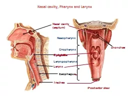
Oropharynx Laryngopharynx Trachea Esophagus Larynx Nasal cavity septum Nasopharynx chonchae Epiglottis Posterior view 307 Larynx Thyroid cartilage Laryngeal ventricle Clemente pl 562 ID: 913130
Download Presentation The PPT/PDF document "Nasal cavity, Pharynx and Larynx" is the property of its rightful owner. Permission is granted to download and print the materials on this web site for personal, non-commercial use only, and to display it on your personal computer provided you do not modify the materials and that you retain all copyright notices contained in the materials. By downloading content from our website, you accept the terms of this agreement.


Slide1
Nasal cavity, Pharynx and Larynx
Oropharynx
Laryngopharynx
Trachea
Esophagus
Larynx
Nasal cavity (septum)
Nasopharynx
chonchae
Epiglottis
Posterior view
Slide2#307 Larynx
Thyroid cartilage
Laryngeal ventricle
Clemente pl. 562
Gray’s p.956
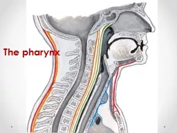
Slide3Slide 307 : larynx
v
ocal fold
thyroid cartilagefalse vocal foldaka vestibular foldepiglottis
(elastic cartilage)chricoid cartilagevocalis m.
Slide4Slide 007 : esophagus & trachea
esophagus
trachea
anteriorneurovascular bundletrachealis muscle
t
rachealis
muscle
Slide5Slide 008: trachea
gc
gland
adventitiasubmucosamucosa
lumen of ?
Slide6Slide 200: trachea & esophagus
Slide7Slide 0027: lung
bronchus
pa
br-iolealveolus
Slide8Slide 0028: lung
*
*
***
Slide9Slide 0028: lung
m
ast cell
type II cellstype I cell
endothelial cellendothelial cell
type I cell
Slide10“Extra Slides” Orientation Images
Slide11UMich
#40
; Trachea
Respiratory epithelium
Basement membrane
Elastic fibers
Slide12Terminal bronchiole
Respiratory bronchiole
Bronchiole
Bronchi
Alveolar sacs and ducts
Slide13UMich
#130-1 Lung
2?
1?
Bronchus
Pulmonary artery
Slide14UMich #
130-1 Lung
Pulmonary artery
Close to bronchial trees relatively thin wall (media) elastic laminae relatively wide lumen
Slide15#130-1 Lung
Bronchial cartilage
4?
3?
Bronchial vein
Bronchial artery
Only in the wall of large bronchi. Ordinary arteries with relatively thick media.
Slide16Bronchial Arteries
Left bronchial arteries
Right bronchial artery
Slide17#130-1 Lung
Ciliated and non-ciliated (Clara) cells No goblet cells Prominent sm. M.
Ciliated cells
non-ciliated cells
Smooth muscle
Bronchiole
Slide18UMich
#
132-2 (Terminal) Bronchiole & Pulmonary arteryBronchiole
P. A.Smooth Muscle
Slide19UMich #
132 Lung
6?
5?
7?
Terminal bronchiole
Respiratory bronchiole
P.A.
Low cells
alveolar pocketing
Knobs of sm. muscle
(arrows)
Slide20UMich #
129 Lung
8?
Alveolar duct
9?
alveoli
Alveolar sac
Slide21UMich
#130-1 Lung
Type I Pneumocyte
Type II Pneumocyte (surfactant)
Macrophage
10?
11?
12?
Slide22UMich #
130-1 Lung
Pulmonary vein
Away from bronchial trees
thin wall
13?
Slide23UMich
#130-2 Lung
Pulmonary
veins
Slide24Intrapulmonary circulation
Pulmonary artery
Pulmonary vein
Pulmonary vein
Slide25UMich #
129 LungArea showing transition from terminal bronchiole
down to alveoli (terminal bronchiole (tb) respiratory bronchiole (rb) alveolar duct (ad) alveolar sacs (as) alveoli (a)
tb
rb
ad
asaadas
asaa