
Amber Earlywine 12 Tyler Kinder 3 Rebecca Dutch PhD 13 1 NSF REU program Lexington KY 2 Berea College Department of Chemistry Berea KY 3 Department of Cellular and Molecular Biochemistry University of Kentucky Lexington KY ID: 919824
Download Presentation The PPT/PDF document "Inhibiting Human Metapneumovirus (HMPV) ..." is the property of its rightful owner. Permission is granted to download and print the materials on this web site for personal, non-commercial use only, and to display it on your personal computer provided you do not modify the materials and that you retain all copyright notices contained in the materials. By downloading content from our website, you accept the terms of this agreement.


Slide1
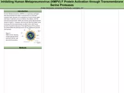
Inhibiting Human Metapneumovirus (HMPV) F Protein Activation through Transmembrane Serine Proteases
Amber Earlywine1,2, Tyler Kinder3, Rebecca Dutch Ph.D1,3 1NSF REU program, Lexington, KY; 2Berea College Department of Chemistry, Berea, KY; 3Department of Cellular and Molecular Biochemistry, University of Kentucky Lexington, KY
Human Metapneumovirus (HMPV) is a respiratory pathogen that was discovered in 2001, within the pneumoviridae family. It has become an important research topic because of its symptoms of severe acute upper and lower respiratory virus in children, the elderly, and the immunocompromised. HMPV has several surface glycoproteins shown in Figure 1. However, the only one that has shown to be necessary for viral transmission to other cells is the Fusion protein (F protein). Figure 2. Cleavage event caused by activation of the F protein by Heparan Sulfate Proteoglycans and a host cell proteaseBecause of the importance of the F protein, it can be determined which proteases bind and cause cleavage. The goal of this project was to test if different proteases such as HAT (TMPRSS11D), TMPRSS2, TMPRSS4, Matriptase, KLK5, or KLK12 cleave the F protein so that a protease inhibitor can be used to stop viral spread. Although the project focused mostly on TMPRSS2 and TMPRSS4.Figure 3. Life cycle of HMPV F protein within host cell
Figure 4. Cell culture process and expected Western Blot AnalysisFigure 5. Western Blot AnalysisTMPRSS2 ran on a 10% gel at 10ug concentration with the HMPV F protein. Trypsin was used as a positive control for the cleavage of the F protein. gure 6. PCR Cloning process to sub-clone the TMPRSS4 insert from a pcDNA plasmid to a pcAGGS plasmid.
Introduction
Methods
Figure 1.
HMPV
virion showing surface glycoproteins such as F, M, SH, and attachment proteins
S
S
F
P
HRA
HRB
TM
F2
F1
-
TM4
pcAGGS
TMPRSS4 Insert
+
F0
F1
Transfection
Lysis
Western blot
a
nalysis demonstrates
p
resence of TMPRSS4 insert after PCR reaction
Figure 7. Western blot of TMPRSS4 insert in varying concentrations
Future Directions
Test expression of
TMPRSS4 in
pcAGGS
after sub-clone and test
if it cleaves HMPV F
Using a protease inhibitor to stop activation of F protein and prevent further viral spread
Using a protease inhibitor as a natural therapeutic for those who are already infected
Conclusion
Based on the Western blot analysis, it can be presumed that TMPRSS2 cleaves the F protein in HMPV and can potentially be inhibited. There is not enough evidence to conclude whether TMPRSS4 cleaves the F protein or not, but future experiments could provide this answer.
References
Acknowledgments
I would like to thank the Dutch Lab, the University of Kentucky NSF REU program, and the Berea College internship office for allowing me to have this internship experience. Also, great thanks to the Dutch Lab for their guidance on this project and to Tyler his mentorship.
Plasmid Cloning by PCR. (n.d.). Retrieved July 18, 2017, from https://www.addgene.org/protocols/pcr-cloning/
https://www.ncbi.nlm.nih.gov/pubmedhealth/PMHT0025751/
RXN Vol. 20 20 50 50
Template DNA 50 250 50 250