
NOMENCLATURE HLA specificities are identified by a letter for locus and a number A1 B5 etc and the haplotypes are identified by individual specificities eg A1 B7 Cw4 DP5 DQ10 DR8 Spe ID: 942299
Download Pdf The PPT/PDF document "MHC MAJOR HISTOCOMPATIBILITY COMPLEX MHC..." is the property of its rightful owner. Permission is granted to download and print the materials on this web site for personal, non-commercial use only, and to display it on your personal computer provided you do not modify the materials and that you retain all copyright notices contained in the materials. By downloading content from our website, you accept the terms of this agreement.


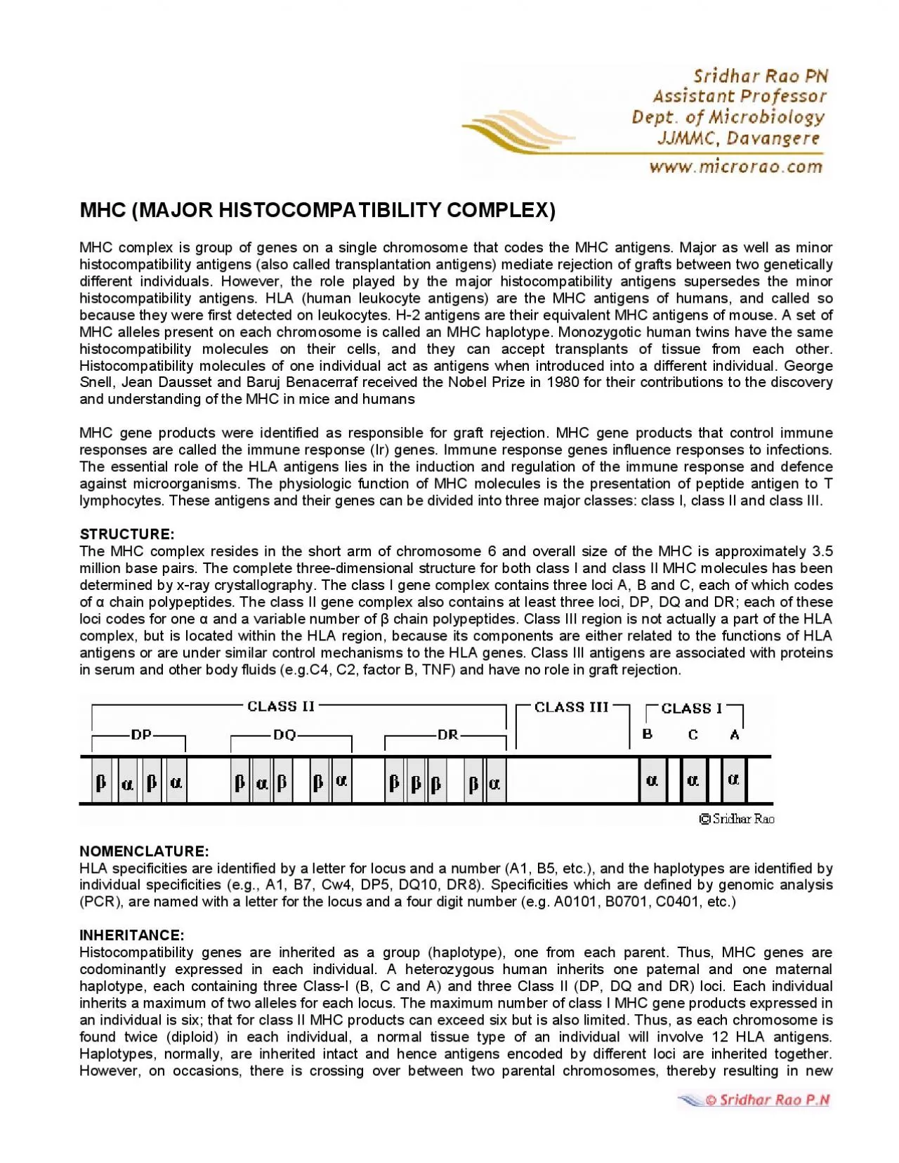
MHC (MAJOR HISTOCOMPATIBILITY COMPLEX) MHC complex is group of genes on a single chromosome that codes the MHC antigens. Major as well as minor histocompatibility antigens (also called transplantation antigens) mediate rejection of grafts between two genetically different individuals. However, the role played by the NOMENCLATURE: HLA specificities are identified by a letter for locus and a number (A1, B5, etc.), and the haplotypes are identified by individual specificities (e.g., A1, B7, Cw4, DP5, DQ10, DR8). Specificities which are defined by genomic analysis recombinant haplotypes. There is no somatic DNA recombination that occurs for antibodies and for the TCR, so the MHC genes lack recombinational mechanisms for generating diversity. Many alleles of each locus permit thousands of possible assortments. There are at least 1000 officially recognized HLA alleles. Class I antigens are expressed on all nucleated cells (except those of the central nervous system) and platelets. The class II antigens are expressed on antigen presenting cells such as B lymphocytes, dendritic cells, macrophages, monocytes, Langerhans cells, endothelial cells and thymic epithelial cells. Cytokines, especially interferon gamma (IFN-), increase the level of expression of class I and class II MHC molecules. MHC CLASS I MOLECULE: Class I MHC molecules contain two separate polypeptide chains, the heavier (44-47 KDa) alpha chain and the lighter (12 KDa) beta chain. The carboxyl end of chain resides inside the cell while the amino end projects on the surface of cell with a short intervening hydrophobic segment traverses the membrane. chain is coded by the MHC genes and has three globular domains 2 and 2-microglobulin is encoded by a gene on another chromosome. The 3 domain is non-covalently associated with the 2 microglobulin. chain and 2-microglobulin are members of the Ig superfamily. Without the 2 microglobulin, the class I antigen will not be expressed on the cells surface. Individuals with defective 2 microglobulin gene do not express any class I antigen and hence they have a deficiency of cytotoxic T cells. A peptide-binding groove is formed between 1 and 2 helices with beta-pleated sheet as its floor. A peptide of 8-10 amino acids long can be presented in this groove. The alloantigenic sites that carry determinants specific to each individual are found in the 1 and 2 domains. The greatest variability in amrphism) occurs in 1 and 2 sequences that line the wall and fl
oor of the groove that binds the peptides. The polymorphism among class I MHC gene products creates variation in the chemical surface of the peptide-binding groove so that various peptide molecules can be accommodated. The specific binding of a peptide molecule in the peptide-binding groove of MHC requires the peptide to have one or more specific amino acid at a fixed position. Such sites are termed anchor sites. The other amino acids can be variable so that each MHC molecule can bind many different peptides. 1 and 2 domains also bind T cell receptor (TCR) of CD8 T lymphocytes. The parts of these domains that are in contact with TCR also show polymorphism. The immunoglobulin-like region of 3 domain is constant (shows no variation) and is non-covalently bound 2 microglobulin. The importance of the highly conserved region of 3 is that CD8 molecules present on CD8 T lymphocytes binds to this region. CD8 T lymphocytes recognizes peptide antigen only when it is presented by the antigen presenting cell in the peptide binding groove of MHC I molecules. Class I molecules present peptide fragments in the cytosol (endogenous antigen, which could be fragments of viral or tumour proteins) to the CD8 lymphocytes. MHC CLASS II MOLECULE: MHC class II molecules comprise two non-identical and non-covalently associated polypeptide chains ( and These two chains have amino ends on the surface, a short transmembrane stretch and intracytoplasmic carboxyl ends. Both chain (34 kDa) and chain (28 kDa) are MHC-encoded and polymorphic. The domains closest to the membrane in each chain are structurally related to immunoglobulins. With the exception of the 1 domain, all domains are stabilized by disulfide bridges. The chain is shorter than the chain and contains the alloantigenic sites. A peptide binding groove is formed in between 1 and 1 domains with a beta pleated floor. As in the case for class I MHC, the greatest polymorphic variability in the amino acids is in those facing the groove. This in turn determines the chemical structure of the groove and influences the specificity and affinity of peptide binding. Peptides associated with class II MHC are 13-25 amino acids long. As with class I MHC, anchor sites for one or more amino acids also exist in the groove of the class II MHC molecule. 2 and 2 are largely non-polymorphic. During antigen presentation, CD4 molecule of Helper T lymphocyte binds to 2 domain of the class II MHC molecules. Exogenous antigens (fragmen
ts of bacterial cells or viruses that are engulfed and processed by antigen presenting cell) are presented to helper T-cells along with MHC II molecules. Because each MHC molecule (I and II) can bind many different peptides, the binding is said to be degenerate. CD 4 Helper T lymphocytes can antigen only when II molecules. CD8 Cytotoxic T lymphocytes can antigen only when molecules. HLA TYPING: Because some HLA antigens are recognised on almost all of the tissues of the body (with few exceptions), the identification of HLA antigens is also described as "Tissue Typing". HLA matching between donor and recipient is desirable for allogenic transplantation. Class I typing methods include test such as microcytoxicity (for typing A, B, C loci) and cellular techniques such as CML (for HLA-DPw typing). Class II typing involves cellular techniques such as MLR/MLC (for DR typing) and molecular techniques such as PCR and direct sequencing (for DR, DQ typing). Serologic methods: Serologic techniques provide one of the most simplest and fastest methods for histocompatibility testing. These methods use sera that contain specific antibodies to HLA antigens. Tissue typing sera for the HLA were obtained in the past, from multiparous women who were exposed to the childs paternal antigens during the parturition and subsequently developed antibodies to these antigens. More recently they are being produced by the monoclonal antibody technology. Microcytotoxicity assay: This is done by exposing the unknown lymphocyte to a battery of antisera of known HLA specificities. Lymphocytes are isolated from the peripheral blood (or from lymph node or spleen in cadavers) and separated from other cells by buoyant density gradient centrifugation. For HLA I antigens, T lymphocytes are chosen while for HLA II antigens, B lymphocytes are chosen. An array of anti-HLA sera covering full range of HLA types are chosen. Individual serum is dispensed into microtitre wells. Approximately 2000 lymphocytes are dispensed per well and incubated. Complement is then added to each well and incubated. The duration of incubation is different for T and B lymphocytes. If the antibodies bind to lymphocytes, complement gets activated and results in lysis of that lymphocyte. The damaged cells are not completely lysed but suffer sufficient membrane damage to allow uptake of vital stains such as eosin Y, Trypan Blue or fluorescent stains such as Ethidium Bromide. Live cells dont stain but the dead c
ells take up the stain. Antibody screening: This is used to detect the presence of HLA antibodies in the potential transplant recipients. A highly sensitive solid phase ELISA is used to detect antibodies in recipients serum. Purified preparations of HLA antigens are adsorbed on the solid phase of plastic plates. Recipients serum is then added to different HLA antigen coated wells. After the removal of unbound antibodies by washing the wells are treated with enzyme-labelled anti-gamma globulin. The wells are washed and treated with colour generating substrate. If the recipient is positive for the HLA type of the donor (that means recipient has antibodies directed against donors antigens), then transplantation is not possible. Cellular Assays: Lymphocytes from one donor, when cultured with lymphocytes from an unrelated donor, are stimulated to proliferate or become cytotoxic. This proliferation is due to a disparity in the class II MHC (DR) antigens. Mixed Leukocyte Reaction (MLR) or Mixed Leukocyte Culture (MLC): T cells of one individual interact with allogeneic class-II MHC antigen bearing cells (eg.B cells) of unrelated individual. When lymphocytes from individuals of different class II haplotypes are cultured together, blast cell transformation and mitosis occurs. The irradiated or mitomycin-C treated stimulator cells of recipient (usually containing B cells, macrophages, dendritic cells) are mixed with CD4 cells of responder (donor). The donor cells respond to different class II antigens on stimulator cells and undergo transformation (DNA synthesis and enlargement) and proliferation (mitogenesis). These changes were recorded by the addition of radioactive (tritiated, 3H) thymidine into the culture and monitoring its incorporation into DNA. Cell mediated lympholysis (CML): The responder cells not only undergoes blast transformation and proliferation on contact with different MHC II molecules, they also give rise to cytotoxic cells. These cytotoxic cells in turn identify the HLA I antigen on the stimulator cells and kill them. Molecular techniques: These methods involve detection of the genes coding for the antigens rather than detecting the antigen itself. These are Sequence-specific PCR, Restriction fragment length polymorphism and sequence specific oligonucleotide probe SIGNIFICANCE OF HLA TYPING: Applications of Applications of Histocompatibility Testing Anthropology: The fact that HLA types vary very widely among different ethnic popu
lations has allowed anthropologists to establish or confirm relationship among populations and migration pattern. HLA-A34, which is present in 78% of Australian Aborigines, has a frequency of less than 1% in both Australian Caucasoids and Chinese. Paternity Testing: If a man and child share a HLA haplotype, then the possibility is there that the man may be the father but not proven. However, if they dont match or share a haplotype then it is agreed that he is Transplantation: Because HLA plays such a dominant role in transplant immunity, pre-transplant histocompatibility testing is very important for organ transplantation. Results with closely related living donors matched with the recipient for one more both haplotypes are superior than those obtained with unrelated cadaveric donors. Transfusion Forensic science Disease Correlation A number of diseases have been found to occur at a higher frequency in individuals with certain MHC haplotypes. Most prominent among these are ankylosing spondylitis (B27), celiac disease (DR3), Reiter's syndrome I. Disease associations with Class I HLA Ankylosing spondylitis (B27), Reiter's disease (B27), Acute anterior Uvietis (B27), Psoriasis vulgaris (Cw6) II. Disease associations with Class II HLA Hashimoto's disease (DR5), Primary myxedema (DR3), Graves thyrotoxicosis (DR3), Insulin-dependent diabetes (DQ2/8), Addison's disease (adrenal) (DR3), Goodpasture's syndrome (DR2), Rheumatoid arthritis (DR4), Juvenile rheumatoid arthritis (DR8), Sjogren's syndrome (DR3), Chronic active hepatitis (DR3), Multiple sclerosis (DR2, DR6), Celiac disease (DR3), Dermatitis herpetiformis (DR3) No definite reason is known for this association. However, several hypotheses have been proposed: antigenic similarity between pathogens and MHC, antigenic hypo- and hyper-responsiveness controlled by the class II genes are included among them. Possible explanation for these associations is that the HLA antigen itself plays a role in disease, by a method similar to one of the following models:- a) by being a poor presenter of a certain viral or bacterial antigen b) by providing a binding site on the surface of the cell for a disease provoking virus or bacterium c) by providing a transport piece for the virus to allow it to enter the cell by having a such a close molecular similarity to the pathogen that the immune system fails to recognise the pathogen as foreign and so fails to mount an immune response against it. Last edited on June