
Zheng Feng Lu PhD Stephen Thomas MD Department of Radiology University of Chicago Chicago IL Question What is the cause of the echogenic band Arrow obscuring part of the urinary bladder ID: 1021879
Download Presentation The PPT/PDF document "Tuesday Case of the Day - Physics" is the property of its rightful owner. Permission is granted to download and print the materials on this web site for personal, non-commercial use only, and to display it on your personal computer provided you do not modify the materials and that you retain all copyright notices contained in the materials. By downloading content from our website, you accept the terms of this agreement.


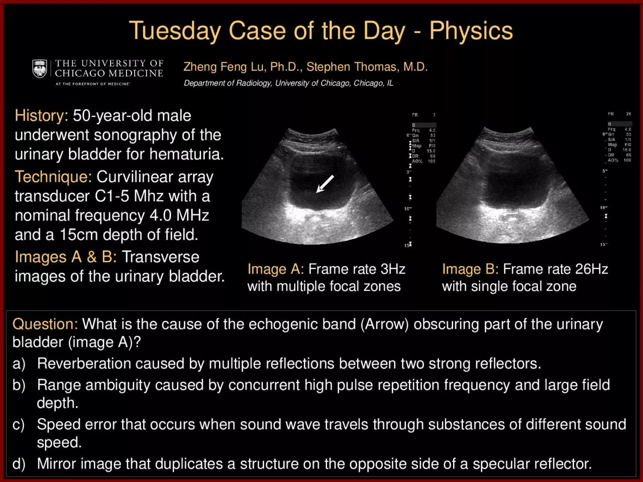
1. Tuesday Case of the Day - PhysicsZheng Feng Lu, Ph.D., Stephen Thomas, M.D.Department of Radiology, University of Chicago, Chicago, ILQuestion: What is the cause of the echogenic band (Arrow) obscuring part of the urinary bladder (image A)?Reverberation caused by multiple reflections between two strong reflectors.Range ambiguity caused by concurrent high pulse repetition frequency and large field depth.Speed error that occurs when sound wave travels through substances of different sound speed.Mirror image that duplicates a structure on the opposite side of a specular reflector.Image A: Frame rate 3Hz with multiple focal zonesImage B: Frame rate 26Hz with single focal zoneHistory: 50-year-old male underwent sonography of the urinary bladder for hematuria. Technique: Curvilinear array transducer C1-5 Mhz with a nominal frequency 4.0 MHz and a 15cm depth of field.Images A & B: Transverse images of the urinary bladder.
2. Diagnosis: The correct answer is (b), range ambiguity (artifact) caused by concurrent high pulse repetition frequency and large field depth.
3. DiscussionA B-mode ultrasound image is formed by emitting a pulse and collecting the resultant echo signal along the insonified path. After each transmit pulse, sufficient time must be allocated for the echoes to return using the following range equation:D = ct/2Where D is the depth, t is the delay time between pulse emission and echo reception and c is the sound propagation speed. Echoes from a deeper depth takes a longer time to return. When echoes from deep structure from the previous transmit pulses are received simultaneously as the echoes from shallow structure insonified by the present pulse, the “range ambiguity” artifact occurs. This typically happens when multiple focal zones are set for a large field-of-view because multiple pulses are needed along the same beam line while each pulse can only focus at one particular depth. Consequently, very high pulse repetition frequency is typically employed for multiple focal zone settings, which causes the “range ambiguity” artifact especially when the field-of-view is large. This may become more obvious when the object has low attenuation such as a urinary bladder. By reducing the number of focal zones to one, the pulse repetition frequency is reduced; thus the range ambiguity artifact is removed.Clinically, range ambiguity artifact is important to recognize and correct as it can obscure important features such as tissue planes and organ pathology.
4. ReferencesRT O’Brien, JA Zagzebski and FA Delaney, “Ultrasound corner: range ambiguity artifact”, Veterinary Radiology & Ultrasound, Vol. 42, No. 6, 2001, pp 542 – 545.A Goldstein, “Range ambiguities in real-time ultrasound”, J Clin Ultrasound, Vol 9, No. 2, 1981, pp83-90.