
Describe the proper technique for performing microscopic urinalysis Identify risk factors that increase the likelihood of finding malignancy during evaluation of hematuria Explain the significance o ID: 959821
Download Pdf The PPT/PDF document "Hematuria Cystoscopy Urine Cytology UTI ..." is the property of its rightful owner. Permission is granted to download and print the materials on this web site for personal, non-commercial use only, and to display it on your personal computer provided you do not modify the materials and that you retain all copyright notices contained in the materials. By downloading content from our website, you accept the terms of this agreement.


Hematuria, Cystoscopy, Urine Cytology, UTI, bladder cancer BJECTIVESAt the end of this clerkship, the learner will be able to: Describe the proper technique for performing microscopic urinalysis. Identify risk factors that increase the likelihood of finding malignancy during evaluation of hematuria. Explain the significance of finding red Identify the indications for screening blood cells in the urine. When visible to the patient, it is termed gross hematuria, while microscopic hematuria is not visible to the naked eye but rather detected by the microscopic examination of the urinary The dipstick method to detect hematuria depends on the ability of hemoglobin to oxidize a chromogen indicator with the degree of the indicator color change proportional to the degree of hematuria. Dipsticks have a sensitivity of 95% and a examination of the urine as patients may test positive when there is not true blood there. Free hemoglobin, myoglobin and certais (povidone- iodine) will give rise to these false positive readresults of the microscopic urinalysis will help differentiate these confounders. The presence of significant proteinuria (2+ or greater) suggests a nephrologic origin for hematuria. The presence of many epithelial cells sugge
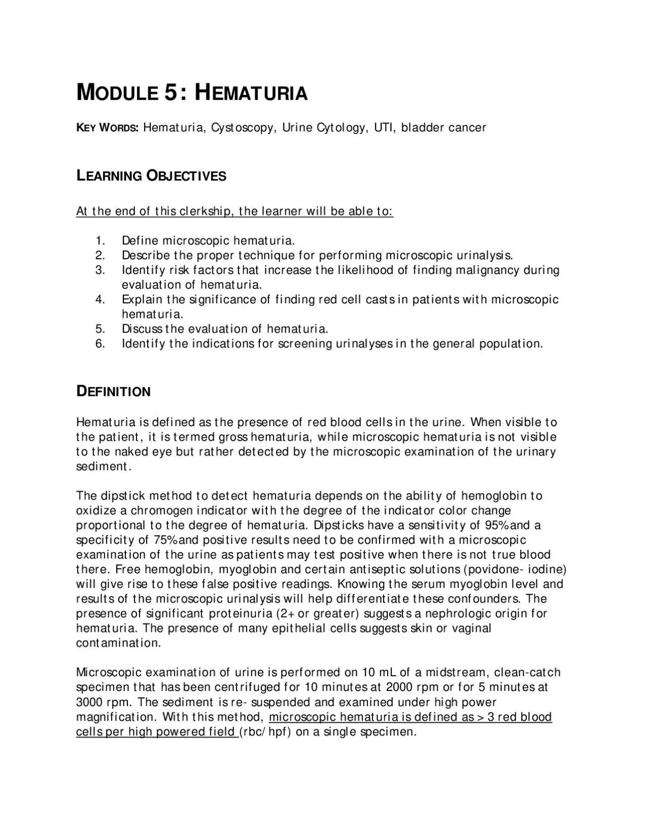
sts skin or vaginal Microscopic examination of urine is performed on 10 mL of a midstream, clean-catch pic hematuria is define�d as 3 red blood cells per high powered field (rbc/hpf) on a single specimen. Figure 1: Red blood cells observed on high power microscopy of urine sediment. The presence of red cell casts, dysmorphic red blood cells, leukocytes, bacteria and crystals should also be includPIDEMIOLOGYa ranges from 1-20% depending on the ing significant urologic disease in these patients also varies with associated risk factors which include: cy in Patients with Hematuria History of cigarette smoking History of chemical exposure (cyclophosrgency, frequency, dysuria) Prior urologic disease or treatment documenting a urologic malignancy in patients referred , no major health organization currently uria in asymptomatic patients. Instead, the decision to obtain a urinalysis (dipstick or microscopic) is based on the interpretation of clinical findings by the evaluating physician. TIOLOGYcan be from anywhere in the urinary tract between the kidney glomerulus and the urethral meatus (Figure 2). Figure 2: Human urinary tract anatomy that is at risk when hematuria is found. From: Nlm.nih.gov Causes of hematuria
may be Glomerular or Nonglomerular. Glomerular causes generally arise from the can be further subdivided by whether the t (kidney and ureter) or lower urinary tract (bladder and urethra) (Figure 2). In general, urologists are concerned with structural and pathologic conditions that are visible on imaging and endoscopic examination whereas glomerular hematuria is Urinary findings suggestive of a glomerular source for the patients hematuria include red cell casts, dysmorphic red blood cells Figure 3: Example of dysmorphic red blood cells consistent with renal or glomerular hematuria. The presence of red cell casts (Figure 4) in the urinary sediment is strong evidence for glomerular hematuria. Figure 4: Example of a red cell cast (arrow) in the urinary sediment. Although protein may enter the urine along with the red blood cells regardless of the renal parenchymal process and should prompt consultation with a nephrologist. The more common causes of glomerular hematuria are listed in Table 2. A more comprehensive list is found in references 2 and 4. IgA nephropathy (Bergers disease) Thin glomerular basement membrane disease Hereditary nephritis (Alports syndrome) cause of asymptomatic glomerular gnificant proteinuria, typica
lly follows a for the condition although fish oils may benefit patients with progressive disease. often classified by location. The more commonly encountered upper and lower urinary tract etiologies are listed in Table 3. Although transitional cell carcinoma involving the urinary bladder is the most common malignancy discovered in patients with asymptomatic microhematuria, a benign process is far more the more likely explantract infection, urinary tract stones and pr Table 3 Common Causes of Non-Glomerular Hematuria Upper Tract Renal cell cancer Transitional cell carcinoma Bacterial cystitis (UTI) Benign prostatic hyperplasia Transitional cell carcinoma Spurious hematuria (e.g. menses) Benign hematuria (e.g. interstitial cystitis, trigonitis) Excessive anticoagulation from oral anticoagt lead to de novo ee and duration of hematube influenced by such therapy. American Urologic Association guidelines suggest urologic and nephrogenic evaluation in patients with hematuria on anticoagulation VALUATIONThe cornerstone of evaluating patients with hematuria is a thorough medical history and directed physical examination. A prior history of urologic disease or interventions is an important feature. Also, the presence)
may suggest an etiology for the patients chemotherapeutic agents, in particular ne, have been associated with hemorrhagic cystitis. Both cigarette smoking and occupational exposures to aniline dyes and aromatic amines used in certain manufacturing procebladder (eg indwelling catheter, stones, and The presence of edema and cardiac arrhythmias may suggest the nephrotic syndrome and atrial fibrillation (with the possibility of renal embolization), respectively. Costovertebral angle tenderness is suggestive of ureteral obstruction, often secondary to stone disease, in the afebrile patient.present the diagnosis of pyelonepIf the patient has not had a formal microscopic urinalysis this should also be part of the initial evaluation. As noted earlier, the di results in patients with myoglobinuria. Also, some patients may present with red of spurious hematuria may reveal a norm in patients with significant urologic In addition to identifying the number of red blood cells per high powered field, the nitrites, etc may suggest infection. If infection is suspected, a confirmatory urinurinalysis performed after the infection has been treated. Patients with findings consistent with glomerular hematuria should be referred to nephrology f
or further including the urinalysis, patients with non- glomerular hematuria may be stratified as high risk or low risk for significant tients with gross hematuria or those with asymptomatic factors noted in Table 1 are considered high risk and should undergo a thorough urologic evaluation. Patients with still warrant a complete evaluation. Bloocomplete blood count, and coagulation parameters can be useful, and PSA may be checked in men dependBecause of the significant diseases that can cause nonglomerular hematuria, a indicated. Imaging studies are used to and ureters) whereas direct endoscopic needed for the lower urinary tract (Figure Figure 5: Flexible cystoscopy is used to examine the lower urinary tract in hematuria cases. Cystoscopy is recommended by the AUA guidelines in all patients at least 35 years of age with microhematuria and in all patients who present with gross hematuria. For patients less than 35 years of age with microhematuria, cystoscopy may be performed at the discretion of the clinician based on the presence of risk factors for malignancy. Currently urine cytology or other tumor markers are not routinely recommended in the evaluation of asymptomatic microhematuria, but may be considered in patients with r
isk factors. Delayed excretory cross-sectional abdominal and pelvic imaging is necessary to evaluate the upper urinary tract and exclude upper tract malignancies. Given its relatively high sensitivity and specificity, CT urography (CTU) is the preferred modality. If renal function or iodine allergies preclude the ability to undergo CTU, then MR urography or retrograde pyelograms with non-contrasted renal imaging can be considered. With this evaluation strategy, a cause for hematuria is identified in roughly 57% of patients with microhematuria and 92% of patients with gross hematuria. Malignancy is identified in approximately 3-5% of patients presenting with microhematuria and 23% of patients presenting with gross hematuhematuria, a urinalysis should be checked annually. Patients with persistent hematuria after a negative initial evaluation warrant repeat evaluation in 3-5 years, Diagnosis, Evaluation and Follow-up of Asymptomatic Microhematuria (AMH) in Barocas DA, Castle EP, Lang EK, Leveillee RJ, Messing EM, Miller SD, Peterson AC, Turk TMT, Weitzel W. American Urological Association (AUA) 2012. Accessible at: Campbell-Walsh Urology, 11th edition, WB Saunders; 2016. Pocket Guide to Urology, 5th edition, (Updated July