

Dr Than Kyaw 27 February 2012 COAGULATION Complex process of blood clotting Important part of hemostasis Cessation of blood loss from damaged vessels Disorders of coagulation can lead to an increased risk of bleeding hemorrhage or clotting thrombosis ID: 909822
Download Presentation The PPT/PDF document "Lab Work 1 Anticoagulants, ESR and PCV" is the property of its rightful owner. Permission is granted to download and print the materials on this web site for personal, non-commercial use only, and to display it on your personal computer provided you do not modify the materials and that you retain all copyright notices contained in the materials. By downloading content from our website, you accept the terms of this agreement.


Slide1
Lab Work 1Anticoagulants, ESR and PCV
Dr Than
Kyaw
27 February 2012
Slide2COAGULATIONComplex process of blood clottingImportant part of hemostasisCessation of blood loss from damaged vessel(s)Disorders of coagulation can lead to an increased risk of bleeding (hemorrhage) or clotting (thrombosis). (Refer to theory) 2
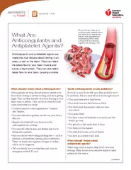
Slide3ANTICOAGULANTSDrugs that prevent the clotting (coagulation) of blood by inhibiting the formation of one or more clotting factorsThe reagents employed should not bring about alteration of blood components. 3
Slide4USES OF ANTICOAGULANTSIn vitro - experiments - blood tests
In vivo - usually given as treatments
- to prevent thrombus extension and embolic complications by reducing the rate of fibrin formation
- Deep vein thrombosis and pulmonary embolism.
-
Myocardial
infarction
-
Cerebrovascular
disease - Blood transfusion
4
Slide55Types of anticoagulants
Ammonium and Potassium
Oxalate
Ammonium oxalate – expand the RBCs
Sodium oxalate – shrink the RBCs
Also k/s double oxalate, balanced oxalate
Mixture of ammonium oxalate and potassium oxalate (3:2)
1
mg of double
oxalate - sufficient
to prevent clotting of 1 ml of blood
action
: precipitate
ionic calcium and
form insoluble
salts
.
safely
used for
haematocrit
,
Hb
estimation
, RBC and WBC counts,
prothrombin
time and
bilirubin
concentration etc.
Slide66Types of anticoagulants
2. Sodium Citrate (
trisodium
citrate)
- 3.8% aqueous solution is used.
- 9 parts of blood: 1 part of sodium citrate solution
-
action
: Chelating
effect on calcium ion
Slide77Types of anticoagulants
3. E.D.T.A (
Versene
or
Sequestrene
)
-
di
-sodium salt of ethylene
diamine
tetra acetic
acid (EDTA); powerful
anti-coagulant
-
action
: stable
chelating effect on calcium
- 1 to 2 mg per ml of blood
- can be used in the blood transfusion
- prevents blood platelets clumping in vitro; so can be employed for
platelets count
- Blood from some birds and reptiles
hemolyzes
when collected
into
EDTA
Slide88Types of anticoagulants
4. Dicoumarol
(
Bishydroxycoumarin
)
- O
btained from spoiled sweet
clover;
inactive in vitro
-
A
ffect
the transport of vitamin K to its site of action.
-
Vit
K is required for normal synthesis of
prothrombin
and
other clotting factors.
-
Interferes
with factor VII and factor X in the liver
- R
educes the Christmas factor (IX)
- Reduces the adhesiveness of platelets
- I
ncreases
antithrombin
concentration in plasma
Slide99Types of anticoagulants5.
Heparin
- a natural anti-coagulant which aids in maintaining blood in a fluid
state.
- main source in the body - mast cells (liver, lungs etc)
- inhibits the reaction between thrombin and fibrinogen
- also interferes with the formation of
thromboplastin
- Heparin retards agglutination and lyses of platelets
- used in the ratio of 0.1 to 0.2 mg per ml of blood
- should not use for blood films; it gives faint blue back-ground on
staining.
Slide1010Types of anticoagulants
6. Hirudin
:
- P
resent in the
buccal
glands of leech
- A
powerful anti-coagulant
- I
nhibits the conversion of
prothrombin
to thrombin.
7.
Venoms
- V
enoms of certain snakes contain a powerful anticoagulant effect
- I
nterferes with the action of
thromboplastin
or
- D
eplete the blood fibrinogen.
Slide11118.
Contact and Smoothness of Surface
-
The clotting of the blood can be prevented when it comes in contact with the tissue fluid or with the substance which is wet or has a smooth surface.
-
When
the vessel in which the blood is collected is coated with thin layer of paraffin wax, blood remains in fluid state for half an hour.
Types of anticoagulants
Slide1212Lab 1. (b)
Erythrocyte sedimentation rate (ESR)
Aim
:
To determine the ESR of the blood sample using
Westergren
/
Wintrobe
tube
Principle
A simple non-specific screening test
I
ndirectly measures the presence of inflammation in the body
A
column of blood taken in a narrow tube
K
eep vertically undisturbed
RBCs tend to settle down due to
higher specific gravity
than plasma
T
endency of RBCs to settle more rapidly in the face of some disease states
U
sually
due to increase
in plasma fibrinogen,
immunoglo-bulins
, and other
acute-phase
reaction proteins
-
Changes in red cell shape or numbers may also affect the ESR.
Slide13Aggregate or Rouleaux formation
Single rouleaux formation
Medium density aggregate
Small high density aggregate
Small
rouleaux
network
Slide1414Method 1
Method 2
Westergren
tube
300 mm in length (graduated 0 – 200 mm)
Internal diameter 2.5 mm
Both ends open
3.8% sodium citrate solution
Blood
Stand (rack)
Wintrobe
tube
110 mm long (graduated 0 – 100 mm)
Internal diameter 3 mm
One end closed
Pasteur pipette
EDTA
Blood
Stand
(rack)
Requirements
:
ESR
Slide15Species
Site of collection and permitted conditions
Dog, Cat
Cephalic
,
saphenous
, femoral and jugular veins
Ruminants
Jugular
vein, tail vein
Swine
Jugular
vein, anterior vena cava, ear veins
Chicken
Brachial
wing vein, right jugular vein
Blood sample collection
ESR
Slide1616ESR
Procedure
(
Westergren
)
Collect 2 ml of blood into a syringe by venue puncture
transfer into a vial containing 0.5 ml of 3.8% sodium citrate solution
(
blood to citrate ratio of 4:1
)
Mix gently the contents of the vial by repeated inversion
Suck the blood mixture carefully into the
Westergren
pipette up to the 0 mark
without introducing any air bubbles.
Fix the pipette vertically in the
Westergren
rack
Allow it to remain for 2 hours
Screw clamp at the top as well as pit at the bottom provided by rubber pads
ensuring right fixing without allowing leakage.
It also avoids the effect of atmospheric pressure.
Observe the pipette for the column of clear plasma at the end of 1 h
and the 2
nd
hour.
Note the readings
in mm per 1
st
hour and 2
nd
hour.
Slide17Slide18ESR
Species
Westergren
tube
Wintrobe
tube
mm at end of 1 hour
mm at end of 2 hour
Record your observed values
after 1 hour and then 2 hour
.
Precautions
The pipettes should be kept perfectly vertical.
Blood column should be without
bubles
.
Use the blood within 2 hours of collection (6 hours if kept 4
C).
The test should be carried out at the room temperature.
Slide1919ESR
Procedure
(
Wintrobe
)
- Performed similarly
as
Westergren
method
- EDTA (or double oxalate)
anticoagulated
blood without extra
diluent
is drawn into the tube.
- The value is usually higher than
Westergren
method.
- The method also is exposed to atmospheric pressure.
Slide20ESR
Some interferences which increase ESR:
increased level of fibrinogen, gamma globulins.
technical factors: tilted ESR tube, high room temperature.
Some interferences which decrease ESR:
abnormally shaped RBC (sickle cells).
technical factors: short ESR tubes, low room temperature, delay in test performance (>2 hours), clotted blood sample, excess anticoagulant, bubbles in tube.
Chronic inflammatory disease (collagen and vascular diseases) increases ESR.
Polycythemia
decreases ESR.
Slide21Red blood cells have settled, leaving plasma at the top of the tube. Reading: 18 mm/hourB
A
A at “0” hr; B after 1 hour
Slide22Lab 1. (c) Hematocrit
or Packed Cell Volume (PCV)
Aim
: to estimate the packed cell volume of the ------------------- blood
Principle
- PCV
(
hematocrit
) - one of the most common blood test performed
-
PCV is the percentage of RBC in the blood.
- E
asy, inexpensive, reliable, and provides valuable information.
-
The term “packed cell volume” describes the principle behind the test.
- B
lood is drawn into a capillary tube,
- Take care not to overfill the tube.
- By c
entrifugation cellular components are ”packed” into the bottom of
the capillary tube.
- RBCs
at the bottom of the tube,
B
uffy coat (leukocytes
a
nd platelets)
C
lear to yellowish top layer is plasma.
Slide23Hematocrit
Procedure
Fill two
hematocrit
tubed
¾ full
with blood containing anticoagulant
(by capillary action).
Blood may be obtained directly from the patient. Use a hematocrit tube containing an anticoagulant. - Wipe the tube with a
kimwipe
or blotting paper
-
Place finger over the “non-blood” end of the tube and push the
opposite end into a clay sealant 3-4 times
- Place the
hematocrit
tube in a centrifuge with the
clay end toward
the periphery
- Place centrifuged
hematocrit
tube on a reader with the top of the clay sealant at the 0% mark and the top of the plasma layer at 100%
Centrifuge at 10000 rpm for 5 minutes.
Slide24Hematocrit
Procedure (continued)
Read the % of RBC which is read at the top of the RBC layer,
do not include the
buffy
coat.
Note and record the color of the plasma.
(
eg
. Clear and transparent, white and cloudy, etc)
Note an increase or decrease in the size of the
buffy
coat.
The
buffy
coat is usually less than 1 mm wide.
Also, note the color of the
buffy
coat which is normally white.
Record your findings carefully and write a lab report.
Slide25Hematocrit
Slide26Format of the report
DVT 1073
Fisiologi
Haiwan
II
Lab work no. ____
Name: ____________ No.
Matrik
: ______________ Date: ________
Title of the laboratory work.
Aim
Requirements
Procedures
Observation and findings
Discussion/Comments/conclusion
Slide27The End