

OUTLINE OF THE TOPIC PART 1 INDICATIONSCONTRAINDICATIONS COMPLICATIONS PART 2 BASIC TECHNIQUEHARDWARE PART 3 CORONARY ANATOMYANGIOGRAPHIC VIEWS PART 4 LESION CHARACTERISTICS HISTORY Werner ID: 908849
Download Presentation The PPT/PDF document "CORONARY ANGIOGRAM -ESSENTIALS" is the property of its rightful owner. Permission is granted to download and print the materials on this web site for personal, non-commercial use only, and to display it on your personal computer provided you do not modify the materials and that you retain all copyright notices contained in the materials. By downloading content from our website, you accept the terms of this agreement.


Slide1
CORONARY ANGIOGRAM -ESSENTIALS
Slide2OUTLINE OF THE TOPIC
PART 1 - INDICATIONS,CONTRAINDICATIONS,
COMPLICATIONS
PART 2 -BASIC TECHNIQUE,HARDWARE
PART 3 -CORONARY ANATOMY,ANGIOGRAPHIC VIEWS
PART 4 -LESION CHARACTERISTICS
Slide3HISTORY
Werner
forsmann
-First angiogram
Cournard
-
Catheterisation
Mason
sones
-coronary angiogram
Andreas
greuntzig
- First angioplasty
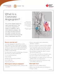
Slide4Slide5Coronary angiogram
Radiographic
visualisation
of coronary arteries after injection of radio opaque contrast media
"Lumen - o - gram"
Cannot
visualise
exterior of the
artery,plaque
and endothelial surface
Does not provide hemodynamic significance of a lesion
Hence the role of FFR,IVUS,OCT
Slide6Types of coronary artery disease
Stable angina - Angina only on exertion
Unstable angina -Angina at
rest,biomarker
neg
NSTEMI -Angina at rest +
Postive
biomarker
-ECG- STD,T inv, No ST elevation
STEMI - ST elevation in > 2
contiguos
leads
Slide7Indications
1.STEMI
2.NSTEMI - High risk
3.Unstable angina -High risk
4.Stable angina -refractory
angina,positive
for inducible ischemia on stress test
5.Valvular heart disease undergoing other cardiac surgery
6.Suspected coronary artery anomalies
Slide8Contraindications
Active infection
Coagulopathy
(Increased
INR,Abnormal
BT,CT)
Severe anemia(hemoglobin <8 mg/dl)
Severe electrolyte imbalance
Active GI bleeding
Slide9Acute aortic valve
endocarditis
Acute renal failure
Known contrast allergy or history of anaphylaxis to contrast agents
Known radiation sensitivity
Caution -
Pregnany
Slide10Complications
1.Acute kidney injury(renal failure)
2.Access site complications
3.Coronary dissection
4.Air embolism
5.Acute pulmonary edema
6.Arrhythmias
7.MI,Stroke
8. Infection
9.Radiation injury(Stochastic effects)
10.Contrast allergy
Slide11Consent
1.Explanation of the procedure
2.Risks involved
3.Risk
vs
clinical benefit
4.Explain to the patient and bystander
5.Written informed consent in patient's own words
6.Reassurance
Slide12A thorough history
Medications
Co
morbidites
In STEMI taken for
primary,quick
history of
drugs,CKD,past
procedures,immunocompr.status
In pts with prior PTCA -
date,indication,hardware
used
In pts with prior CABG -
grafts,arterial
or venous
Slide13Patient preparation
Sedation overnight to allay anxiety
Preparation of parts
Examine access site
Peripheral pulses
Pallor,vitals
Cardiovascular status
Pre procedure
ECG,ECHO,biomarkers,RFT
Slide14During procedure
Constant monitoring of ECG,HR,BP,RR,SPO2
IV line should be ready before procedure
Allen's test on both
hands,Barbeau
method
ECHO
Emergency medications
Intubation set
Defibrillator
Slide15CORONARY CATHETERS
Diagnostic
Thicker shaft
Internal DM
Smaller
Tapering tip
Less Reinforced construction ( 2 layers)
GUIDE
Thinner shaft
Internal DM larger
Non tapering tip
More Reinforced construction ( 3 layers)
Slide16CHARACTERISTICS OF
AN IDEAL CATHETER
Better torque control
Pushability
Flexibility
Trackability
Radio-opacity
Atraumatic
tip
Kink resistance
Slide17Slide18A) TIP LENGTH –
Increased length offers more
stability in target vessel
B) PRIMARY CURVE –
angle of the target vessel
from its parent artery.
C) SECONDARY CURVE --
width of the parent
vessel.
D) TERTIARY CURVE –
normal curvature of the
parent vessel.
E) CATHETER LENGTH –
Usually 100 or 110 cm
Slide19Slide20SIZE MEASUREMENT
FRENCH CATHETER SCALE:
outer diameter of cylindrical medical instruments.
D(mm) = Fr/3
Most commonly in adults -- Diagnostic Catheters of 5 – 7 Fr size.
Colour
coding
Slide21CATHETER MATERIALS
TEFLON
POLYETHYLENE
POLYURETHANE
POLY VINYL CHLORIDE
Slide22Commonly used catheters
Slide23JUDKINS
Curve length = distance between P
(primary curve) & S (secondary curve)
Slide24AMPLATZ
Slide25Slide26MULTIPURPOSE CATHETER
Polyurethane catheter
Single curve with straight tip an end hole and two side holes.
Use: CAG – Both native vessel and graft ,
Ventriculography
, Right heart catheterization.
MP A-1 : 1 end hole only
MP A-2 : 2side holes ,1end hole
MP B-1 : 1 end hole only
MP B-2: 2
sideholes
and an end hole
Slide27Slide28BYPASS CATHETERS
RCB
Resembles JR4 with a tip curved >90 degree
LCB
more secondary curve
Slide29Internal mammary catheter
Same as JR4 except for
tighter primary curve (80degree) and
longer tip
Slide30ACCESS SITE
Puncture site ,
preparation,LA
Femoral,radial
Modified
seldinger
technique
High
puncture,low
puncture -problems
Heparin,spasmolytics
in radial
Slide31ADVANCING THE GUIDEWIRE
Under
flouroscopy
Resistance
At aortic bifurcation - PVOD
Aortic arch
Anomalous
subclavian
in radial
Use of hydrophilic wire
Slide32RADIAL APPROACH
0.035 inch
guidewire
J tip wires - may cause vasospasm
Angled-tip hydrophilic
guidewires
useful to negotiate
anomolous
SCA
Significant
subclavian
tortuosity
can be negotiated by use of a stiff shaft hydrophilic-coated
guidewire
. Having the patient take a deep breath can also straighten the vessel.
Slide33Insertion and Flushing of
the Coronary Catheter
The Catheter with
guidewire
inserted
upto
ascending aorta
Aspirate blood and column made air free
Record baseline tip pressure –
Catheter lumen filled with contrast & look for alteration in tip pressure
Selective engagement of coronary.
Slide34Damping and
Ventricularization
of
the Pressure Waveform
A fall in overall catheter tip pressure (damping) or a fall in diastolic pressure only (
ventricularization
)
Indicates obstruction of the catheter tip or interference with coronary inflow
Slide35Small vessel
Ostial
Spasm
Selective Engagement Of The
Conus
Branch
Ostial
Stenosis
Slide36Cannulation
of the Left CoronaryOstium
Slide37Cannulation
of the Right CoronaryOstium
Slide38Saphenous
Vein and
Arterial Grafts
Right Grafts
– Primary choice - MP
– Alternative – JR , RCB , AL
Left Grafts :
– Primary Choice – JR4 , AL1
– Upward trajectory may require - special LCB , IMA
– More anterior origin – AL
Slide39INJECTION TECHNIQUE
IDEAL
- contrast at an adequate rate and volume to transiently replace the blood contained in the involved vessel with slight but continuous reflux into the aortic root.
VIGOROUS
- coronary dissection or excessive myocardial blushing.
PROLONGED
– increased myocardial
depression,staining
,
bradycardia
The rate and volume of injection - an average 7
mL
at 2.1 ml/second in the left and 4.8
mL
at 1.7 ml/second in the right coronary
Slide40Slide41RCA Branches
Conus
branch
SA nodal branch
RV branch
Atrial
branch
Acute marginal branch
AV nodal branch
Posterior descending artery
Posterolateral
vessel
(branch -PLV/PLB)
Slide42Slide43SURGICAL SEGMENTS OF LAD
Proximal
Ostium
to 1
st
major
septal
perforator or 1
st
diagonal artery whichever is first
Mid
1
st
perforator to 2
nd
diagonal
Distal
D2 to end
Slide44ANGIOGRAPHIC CLASSIFICATION OF LAD
Type 1
Small vessel reaches only 2/3
rd
of way from base of heart to apex
Type 2
reaches the apex of
LV
Type 3
Extends from base to apex &
wraps around the diaphragmatic surface of LV.
Slide45SURGICAL SEGMENTS OF RCA
Proximal
Ostium
to 1
st
main RV branch
Mid
1
st
RV branch to acute marginal branch
Distal
Acute margin to the crux
Slide46LCX
Proximal
Ostium
to 1
st
major obtuse marginal branch
Mid
OM1 to OM2
Distal
OM2 to end
Slide47CORONARY DOMINANCE
Gives rise to PDA,
posterolateral
branch and AV nodal branch
Right dominant circulation: 85%
Left dominant circulation : 8%
Balanced dominant circulation: 7%
RCA give rise to PDA and terminates
LCx
give rise to all PLV and sometimes a parallel posterior descending branch supplying part of the IVS
Slide48ANGIOGRAPHIC VIEWS
Slide49TWO TERMS
The
angulation
defined using two terms .
The first term
- Rotation , the term RAO designates a view where the image intensifier is positioned over the patient's right anterior chest wall.
The second term
- Skew, the amount of
angulation
toward the patient's head (cranial) or foot (caudal) depends on image intensifier position.
Slide50Slide51RIGHT AND LEFT
Slide52CAUDAL AND CRANIAL
Slide53LAO CRANIAL
Slide54Atrioventricular
and Interventricular Planes
Slide55Slide56RAO views
RAO-caudal projection (0- 10° RAO and 15-20° caudal) -
left main bifurcation, the proximal LAD, and the proximal to mid LCX.
Slide57Slide58Slide59RAO/Caudal
Slide60RAO CRANIAL
Shallow RAO-cranial projection (0-10° RAO and 25-40° cranial) –
mid & distal LAD, with origins of the
septal
and diagonal branches
distal RCA or distal LCX.
Slide61RAO CRANIAL
Slide62RAO CRANIAL -RCA
Slide63LAO CRANIAL
Slide64SHALLOW LAO CRANIAL
Slide65Slide66LAO CAUDAL(SPIDER VIEW)
Slide67LAO - CRANIAL
Slide68STEEP LAO CRANIAL
(50 to 60 degrees) LAO and angulated cranial (20 to 40 degrees) skews.
Slide69Left coronary artery
1.
RAO-caudal
- left main, proximal LAD and proximal circumflex
2
.RAO-cranial
- mid and distal LAD without overlap of
septal
or diagonal branches
3.
LAO-cranial
- mid and distal LAD in an orthogonal projection.
4
.LAO-caudal
- left main , proximal circumflex & LAD.
5.AP Caudal & cranial
- LMCA
Slide70RCA
In the LAO projection - appears like a letter C & in RAO position- like a letter L.
To check the correct alignment of the catheter with the
ostial
RCA, the best view is the RAO-shallow cranial projection.
Slide71RIGHT CORONARY ARTERY
1. LAO 60 - Proximal and mid RCA
2. LAO cranial - Distal RCA and its PDA & PLV branches
3. RAO-cranial - PDA & PLV
4. Lateral - Mid-RCA
Slide72Slide73HOW TO DIFFERENTIATE SEPTAL & DIAGONAL
DIAGONALS
The diagonals would be in the left of the screen .
Diagonals move (buckle) during systole.
Branches at 45 degree
SEPTALS
Septals
- the right of the LAD
Septals
are straighter and move very little with ventricular contraction
Branches at 90 degree
**
Exposure of the high diagonal -
a good spider view with steep caudal
angulation
is most likely the best view to expose a high diagonal
Slide74LESION QUANTIFICATION
Slide75CAD is defined as a > 50% diameter
stenosis
in one or more of these vessels
Subcritical
stenoses
< 50%
nonobstructive
CAD.
Obstructive CAD is classified as one-, two-, or three-vessel disease.
Slide76Slide77Slide78Slide79Slide80Slide81Slide82Slide83LESION ARRANGEMENT
Tandem
- two lesions located within one balloon length
Sequential
- two lesions located at a distance longer than the balloon
Slide84TORTUOSITY AND ANGULATION
PROXIMAL VESSEL TORTOUSITY
• Number of >75º bends to reach the lesion
- None
- Mild - one bends
- Moderate - two bends
- Severe - ≥ three bends
LESION ANGULATION
- None/Mild - lesion located on a straight segment or a bend <45º
- Moderate; 45º~90º bend
- Severe; bend >90º
Slide85CALCIFICATIONS
None
Mild
- densities noted only after contrast injection
Moderate
- densities noted only with cardiac motion prior to contrast injection
Severe
-
radiopacities
noted without cardiac motion prior to contrast injection
Slide86OSTIAL LESION
Origin of the lesion ≤ 3mm of the vessel origin
-
Aorto-ostial
-
(
LMCA,
Prox
RCA)
- Branch-
ostial
- major
epicardial
artery
LAD &
LCx
os
Dx
os
OM
os
PDA and PL
os
Slide87Slide88Slide89SYNTAX SCORE
Each lesion is assigned a numerical number and then sum of all lesions score for a patient is calculated to come up with the final numerical SYNTAX score
Patients are divided in 3 groups
:
Low <22
Intermediate 23-32
High >32
Slide90MYOCARDIAL BRIDGE
Myocardial Bridges - coronary arteries occasionally dip below the
epicardial
surface under small strips of myocardium.
During systole, the segment of the artery surrounded by myocardium is narrowed and appears as a localized
stenosis
Most common in LAD
Slide91LAD
Slide92THANK YOU