
Berl 1989 97339346 mitotic nondisjunction in faba Chinese hamster cells Rizzoni 1 C aterina Tanzarella 3 Bianca Gustavino 1 Francesca Degrassi 2 Almerinda Guarino 3 and Eleonora Vitaglian ID: 945694
Download Pdf The PPT/PDF document "SpringerVerlag 1989" is the property of its rightful owner. Permission is granted to download and print the materials on this web site for personal, non-commercial use only, and to display it on your personal computer provided you do not modify the materials and that you retain all copyright notices contained in the materials. By downloading content from our website, you accept the terms of this agreement.


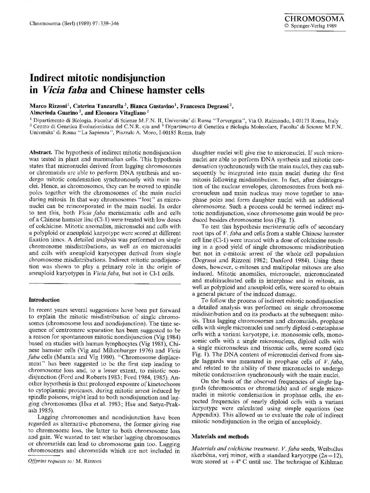
(Berl) (1989) 97:339-346 Springer-Verlag 1989 mitotic nondisjunction in faba Chinese hamster cells Rizzoni 1, C aterina Tanzarella 3, Bianca Gustavino 1, Francesca Degrassi 2, Almerinda Guarino 3, and Eleonora Vitagliano 2 Dipartimento di Biologia, Facolta' di Scienze M.F.N. II, Universita' di Roma "Torvergata", Via O. Raimondo, 1-00173 Roma, Italy 2 Centro di Genetica Evoluzionistica del C.N.R. c/o and 3 Dipartimento di Genetica e Biologia Molecolare, Facolta' di Scienze M.F.N. Universita' di Roma "La Sapienza", Piazzale A. Moro, 1-00185 Roma, Italy hypothesis of indirect mitotic nondisjunction was tested in plant and mammalian recent years several suggestions have been put forward to explain the mitotic misdistribution of single chromo- somes (chromosome loss and nondisjunction). The time se- quence of centromere separation has been suggested to be a reason for spontaneous mitotic nondisjunction (Vig 1984) based on studies with human lymphocytes (Vig 1981), Chi- nese hamster cells requests to: Rizzoni daughter nuclei will give rise to micronuclei. If such micro- nuclei are able to perform DNA synthesis and mitotic con- densation synchronously with the main nuclei, they can sub- sequently be integrated into main nuclei during the first mitosis following misdistribution. In fact, after disintegra- tion of the nuclear envelopes, chromosomes from both mi- cronucleus and main nucleus may move together to ana- phase poles and form daughter nuclei with an additional chromosome. Such a and methods and colehicine treatment. V. faba Weibullus akerb6na, varj minor, with a standard karyotype (2n = 12), were stored at + 4 ~ C until use. The technique of Kihlman DAUGHTER CELLS FIRST MITOSIS AFTER CHROMOSOME IN INTERPHASE (G1) MISDISTRIBUTION MISDISTRIBUTION I ~ 2n-1 2"-' r g i.ed ~ ~ 2n 2n -I+MN (1-syc) 2n-I+MN L/ i IL~_ 2 n 2n+MN (1-syc) 2n+MN 2n-I+MN (1-syc) 1. Scheme for indirect mitotic nondisjunction by the reintegra- tion into the main nucleus of a whole chromosome or chromatid "lost" in a micronucleus. The probabilities of the events involved in the production of variant karyotypes are given in parentheses. Micronuclei which are not mitotically condensed are represented as circles the first mitosis after misdistribution. cronucleus; of chromosome loss; of chromatid loss; of synchronous condensation of micronuclei with main nuclei (1975) was used for germination. Plants were treated for 24 h with 3 x 10⁻⁵ M colchicine (Merck) when roots were 2-4 cm long, immersing roots in the colchicine solution. Colchicine was removed and the roots were washed. Treated root tips were fixed 24, 44 and 64 h after the start of the treatment in a 3/1 methanol/acetic acid mixture. Un- treated roots were fixed at 24 h. For each fixation time, 0.05% colchicine was added to half the samples from both control and treated roots 2 h before fixation to obtain c- metaphases. All root tips were Feulgen stained and squashed. The preparations were mounted in Canada bal- sam. CI-I cells (modal chromosome number s = 21) were cul- tivated on cover slips in Ham's F- 0 medium supplemented with 10% foetal calf serum. Exponentially growing cells were treated with 10-7 M colchicine (Merck) for 24 h; col- chicine was then removed and cells were washed with and reincubated in culture medium. The cells were fixed with 3/1 acid mixture 24, 48, and 72 h after the beginning of the treatment. Untreated control cultures were fixed at 24 h. For each fixation time 5 x 10 -5 M col- chicine was added to half the samples from both control and treated cultures 3 h before fixation to obtain c-meta- phases. A 20 min hypotonic treatment with a 3/1 mixture was carried out on t
hese samples before fixation. Coverslips were stained with Giemsa and mounted in Canada balsam. of 2/3 faba) 1/2 (CI-1) of late telophase nuclei were chosen as borderline values to differentiate between micronuclei and small nuclei, taking into account the number and size of chromosomes of the two cell systems. Cells with two or more nuclei (with or without micronuclei) were classified as multinucleated cells. Cells with one or more micronuclei were classified as micro- nucleated cells. Micronuclei with a prophase appearance were classified as mitotically condensed. Two thousand interphases were analysed for micronu- clei, micronucleated and multinucleated cells, 200 pro- phases for micronuclei with or without mitotic condensa- tion and for synchronous or asynchronous multinucleated cells, 200 metaphases for micronuclei, 200 anaphases for mitotic anomalies and micronuclei, and 200 c-metaphases for polyploid and aneuploid cells. Single lagging chromosomes or chromatids were scored in 1000 ana/telophases 24 h after the beginning of treat- ments. Micronuclei with or without mitotic condensation in 50 single micronucleated prophase cells and different classes of nearly diploid cells with a variant karyotype in 1000 c-metaphases were scored 24h (C1-1) and 44h the start of treatment. These fixation times were chosen so as to analyse the cells in their second mitoses after the start of treatment. At these times the highest fre- quencies of tetraploid, hypotetraploid and micronucleated mitoses were observed, while octoploid and hypooctoploid mitoses were not found. Metacentric (M) and subtelocentric (St) chromosomes can be easily distinguished in faba, haploid karyotype being composed of five subtelocentric chromosomes of sim- ilar size and shape, and one large metacentric chromosome (see Kihlman 1975). Therefore we determined which type of chromosome was involved in misdistribution and in monosomy or trisomy. content of micronuclei in prophase cells of V.faba. DNA content of 50 single micronuclei in nearly diploid prophase cells of faba, at 44 h, was measured with a Zeiss III Photomicroscope equipped with a 01 photome- ter. These micronuclei were classified as pycnotic (homo- geneously stained, presumably dead or degenerating; see McLeish 1954), granular (showing a staining pattern similar to that of the main interphase nucleus; see McLeish 1954) or in mitotically condensed. The 4C value (DNA content of a G2 nucleus) was determined by measuring 20 2n pro- metaphases in the neighbourhood of the measured micro- nuclei. The standard error of the mean 4C value was about 3%. Repeated measurements of the same micronucleus showed a variation range of about 9% of the average value. Nearly diploid prophase main nuclei were accepted within a range between (4 C- 20 %) and (4 C + 3 %) to include both 2n and (2n- 1) nuclei. The upper limit for accepting micro- nuclei was 34% of 4C (corresponding to two metacentric chromosomes). The limits for both main nuclei and micro- nuclei were chosen so that the products of single chromo- some misdistribution only were studied. The DNA content of single chromosomes was measured in five 2n c-meta- phases. expected after a treatment with a low dose of colchi- chine, c-mitoses, and multipolar and "disrupted" (see Ta- Table i. Frequencies of mitotic anomalies in ana/telophases of Viciafaba and CI-1 cells following a 24h treatment with colchicine Fixation c-anaphases Multipolar/ Single lagging time (h) (%) disrupted a chromosomes/ ana/telophases chromatids (%) (%) V. faba Control 0 0 0 24 13_5 9.5 6 44 7 15.5 7 64 4.5 8 3.5 CM cells Control 0 4 0 24 2 10 7 48 3 5 2 72 1 14 6 Each sample consisted of 200 cells. C
olchicine concentration was 3 x 10 -s M for V.faba and 10 -7 M for CI-1 cells " '~ ana/telophases, those with multiple laggards, with or without multipolar spindles ble 1 for definition) mitoses were observed in both cell types together with single lagging chromosomes or chromatids (Table 1) (Ostergren and Levan 1943; Deysson 1968; Gus- tavino et al. 1987). Multinucleated and multiply micronu- cleated cells, derived from c-mitoses and multipolar/dis- rupted mitoses, were observed together with singly micro- nucleated cells, which arise mainly from single lagging chro- mosomes or chromatids (Stubblefield 1964; Kato and Sandberg 1967) (Table 2). Polyploid cells, derived from c- mitoses, and multiply aneuploid cells, derived from multi- polar/disrupted mitoses, (Deysson 1968; Hsu et al. 1983; Satya-Prakash et al. 1984) were observed together with monosomic and trisomic cells, resulting from single chro- mosome misdistribution (Table 3). The ratio between the frequencies of single lagging chro- mosomes or chromatids in ana/telophases (Table 1) and of singly micronucleated cells in interphases (Table 2) is close to 2:1, indicating that most cells passed a mitosis and that each single laggard gave rise to a single micronucleus in one of the daughter cells. The frequency of mitotic anomalies decreased with time, yet it never reached zero. Mitotically condensed micronuclei were only observed in prophase cells (Table 2, Fig. 2b, h). Neither premature chromosome condensation (PCC) (Stubblefield 1964; Kato and Sandberg 1967; Obe and Beek 1982) in mitotic cells nor delayed mitotic condensation in metaplaase, anaphase, teiophase and interphase ceIIs was observed for micronuclei in either cell system. Single lagging chromatids were more frequent than sin- gle lagging chromosomes in both V.faba (ratio close to 6:1) and CI-1 cells (close to 4:3) (Table 4, Fig. 2a, g). On the other hand, only lagging chromosomes were found in Don cells treated with diazepam (Hsu et al. 1983). A ratio close to 5:1 was found between subtelocentric and meta- centric lagging chromosomes or chromatids in V.faba (Ta- ble 4). This ratio is expected assuming a random involve- ment of each chromosome in misdistribution. The frequency of single micronuclei in synchronous mi- totic condensation with main nuclei in prophase was 30% Table 2. Frequencies of micronuclei, and micronucleate and multinucleate cells of Vicia faba and C1-1 following a 24 h-treatment with colchicine" Fixation Interphases b time (h) Singly Multiply Total Multinucleated micronucleated micronucleated micronuelei ceils cells cells (%) (%) (%) (%) Prophases b Metaphases b Ana/ telophases b Micronuclei Multinucleated Micronuclei Micronuclei (%) cells (%) (%) (%) V. faba Control 0.05 0 0.05 0 0 0 0 0 24 1.65 o.os ~.9 o.s5 0.5 (0) ~ 0 0 0 44 2.7 0.25 3.95 1.4 3.5 (1) 3.5 (1.5) a 3 (0) c 3 (0) c 64 3.25 0.3 4.3 1.65 4.5 (1.5) 5 (3.5) 4 (0) 4.5 (0) C1-1 cells Control 0.5 0 0.5 0.3 0 0 0 0.5 (0) 24 1.7 0.3 2.45 0.3 5 (4) 0 1 (0) 0 48 3.2 0.4 4.15 1.05 2 (1) 2 (2) 2(0) 0 72 1.8 0 1.8 0 5 (5) 0 1 (0) 0 a Concentrations of colchicine as in Table 1 b Numbers analysed were 2000 for interphase and 200 each for prophase, metaphase and ana/telophase cells c Numbers in parentheses represent percentages of mitotically condensed micronuclei a Numbers in parentheses represent percentages of synchronous multinucleated cells 3. Frequencies of polyploid and aneuploid cells following a 24 h treatment of Viciafaba and C1-1 cells with colchicine a V. faba Fixation (2n-1) (2n-1) 2n (2n+1) (2n + 2)-(4n- 1) 4n &#x 000;4n time (h) (%) (%) (%) (%) (%) (%) (%) Total micronucleated c-metaphases 4 1 95 0 0 0 0 24 5 1.5 92.5 0 0.
5 0.5 0 44 4.5(1.5) b 4 (1) 76 (0.5) 0 5.5(2) 10 0 64 4.5 3 (1.5) 77.5 (1) 1 5.5(1) 6.5 2(0.5) CI-1 cells Fixation ()c (s-l) s (s+l) (s+2)-(2s- 1) 2s &#xs-l ;2s time (h) (%) (%) (%) (%) (%) (%) (%) Total micronucleated c-metaphases 0.5 6 83 7.5 3 0 0 24 4 13.5 53.5 22 5 2 0 48 11.5 10 (0.5) 50 18.5 7 2.5 0.5 72 1 8 52.5 (0.5) 26 5 5.5 2 0 0 0.5 0.5 Each sample consisted of 200 c-metaphases a Colchicine concentrations as in Table 1 b Numbers in parentheses represent percentages of micronuclei not mitotically condensed c s Modal chromosome number of CI-1 cells Table 4. Analysis of the frequencies of single lagging chromosomes/chromatids and of their products at the first mitosis after misdistribution in colchicine-treated Viciafaba and CI-1 cells- V. faba Number of Number of Number of near 2n cells with a variant karyotype Z z P single chromosome sychronously in 1000 c-metaphases misdistributions condensed in 1000 ana/telophases micronuclei in 50 singly micronucleated prophase cells Lagging Lagging chromo- chromatids somcs M St M St M St (2n--M) (2n--St) (2n-M+MN)(2n-St+MN)(2n+MN) (2n+M) (2n+St) 1 7 8 42 8 7 4 18 0 12 10 3 4 1.498 0.8~0.9 (4.2) ~ (22.3) (0) (11.76) (8.75) (2.5) (2.24) C1-1 cells Number of Number of Number of near s cells with a variant karyotype Z z P single chromosome sychronously in 1000 c-metaphases misdistributions condensed in 1000 ana/telophases micronuclei in 50 singly Lagging Lagging micronucleated (s- 1) (s+ 1) chromo- chromatids prophase cells somes (s- 1 + MN)/(s+ MN) 29 40 34 141 4 204 212.38 (84.5) (11) (91.6) M Metacentric; St subtelocentric; MN micronucleus; s modal chromosome number in CI-I cells. Colchicine concentrations as in Table 1. Lagging chromosomes/chromatids were scored at 24 h after the beginning of the treatment for both systems; micronuclei in prophase cells and near 2n/s cells with a variant karyotype were scored at 44 h in Vicia faba and at 24 h in C1-1 (see Materials and methods). The values for synchronously condensed mieronuclei in prophase cells were taken from Figure 3 for V.faba and were classified as containing metacentric or subtelocentric chromosomes on the basis of DNA amount (see Fig. 3). Chi square values were calculated by pooling the classes with low expected frequencies and computing together the other ploidy classes not described in this table. a The expected frequencies of the different classes of near 2n/s cells with a variant karyotype are given in parentheses and calculated on the basis of the equations described in Appendix 15 t 44 h a 24 h 3 x GI nucleus). Grey areas, white areas, cross-hatched areas, the whole ric (M) c A d A e A c-metaphase lacking f A + 1 CI-1. g A h A i A the basis 1 : 1 (8/50) is close to chromatids and 1 ; close to with an 1 + The observed frequencies of (s- 1) and especially (s+ 1) cells in CI-1 were much higher than those expected (Table 4; see also Danford 1984). data strongly suggest that lagging chromosomes or chromatids and indirect mitotic nondisjunction play a pri- mary role in giving rise to aneuploid karyotypes following single chromosome misdistribution in V. faba, while direct nondisjunction is a rare event. Moreover, while nearly all metacentric chromosomes are regained by main nuclei, about one-sixth of subtelocentric ones are regained. In contrast direct nondisjunction is likely to play a ma- jor role in inducing aneuploid karyotypes in C1-1 cells. The real occurrence of indirect mitotic nondisjunction cannot be verified in this cell type, due to the high incidence of direct nondisjunction. To account for the difference observed between V. faba and C1-1 cells consideration could be made of the forces acting in dis
tribution or misdistribution of chromosomes during mitosis (forces moving kinetochores towards the poles, the association between sister centromeres) in plant and animal cells. ParticL~lar attention should be giver~ to the fate of micronuclei. Reports in the literature on replica- tion and mitotic condensation of micronuclei have mainly been concerned with micronuclei derived from acentric fragments: in cultured mammalian cells an asynchronous pattern of DNA synthesis has been shown, (Kato and Sand- berg 1968; Ikeuchi 1973; Griffiths and Carpenter 1979) which has been linked to PCC. In human lymphocytes both asynchronous (Obe and Beck 1975) and synchronous (Pincu et al. 1985) DNA synthesis pattern have been found. In V.faba DNA replication of micronuclei has been shown to occur either synchronously with main nuclei or not at all (Das 1962; Scott and Evans 1964). In this cell system micronuclei were found to undergo "normal" mitotic con- densation or to remain without mitotic condensation in an interphase state (McLeish 1954; Scott and Evans 1964). Also, PCC in G2 of micronuclei containing whole chromo- somes has been found in mammalian cells (Obe and Beek 1975; Raman 1980; for review see Obe and Beck 1982). However, a high degree of synchrony has been found among micronuclei both in DNA synthesis and in mitosis in PtK-I cells (Ghosh and Paweletz 1984). In summary micronuclear PCC is rather common in mammalian cells, while it is very rare in plant cells (see Obe and Beek 1982). Our data in V.faba show that micronuclei containing single chromosomes are derived mostly from chromatids; some micronuclei replicate before cells enter mitosis and undergo mitotic condensation synchronously with main nu- clei. In contrast, micronuclei which have not completed DNA synthesis cannot undergo mitotic condensation. Some aspects of the process of indirect mitotic non- disjunction should be mentioned: 1. Indirect mitotic nondisjunction gives rise to both (2n + 1) and (2n-1) cells following single lagging chromosomes or chromatids, although (2n + 1) cells are less frequent because not all micronuclei are regained. 2. Micronuclei with more than one chromosome or nuclei belonging to multinucleated cells could be regained follow- ing the same pathway as described for single chromosome misdistribution, but this cannot be demonstrated unambig- uously. Indirect evidence for this is provided by the high degree of synchrony in multinucleated prophase cells (Ta- ble 2). Thus, multiple aneuploidies can appear at the first mitosis after misdistribution by synchronous mitotic con- densation of different nuclei and/or by asynchrony and loss (perhaps through PCC; see Stubblefield 1964; Kato and Sandberg 1967) of uncondensed nuclei (Rainaldi etal. 1987). Recently, selective chromosome replication has been suggested to be the reason for the appearance of hypote- traploid aneuploids from multinucleated cells following c- mitosis (Frackowiak et al. 1986). 3. Different chromosomes in micronuclei might have differ- ent probabilities of being regained by main nuclei. Our data show that in V.faba this probability is close to 1 for the metacentric chromosome and to 1/6 on average, for the subtelocentric chromosomes. A role of nucleolar organizer regions (NORs) within micronuclei in allowing synchro- nous mitotic condensation might explain this result, as NORs are located on the metacentric chromosome of V.faba (McLeish 1954; Das 1962; Scott and Evans 1964). 4. In the absence of micronuclei, aneuploid karyotypes are selected according to their genetic imbalance. During the time between micronucleus formation and its elimination or integration, selection on the "divided karyo
type" may take place at the cellular level, as one chromosome is in the micronucleus and the others are in the main nucleus. It can be asked whether there is a functional integration between the micronucleus and main nucleus, i.e. whether a (2n-1 + MN) cell acts as a (2n-1) cell or as a (2n)/(2n + 1) cell. At the same time, selection on the micronucleus chromosome also takes place (see point 3). Divided karyo- type and micronucleus chromosome selections act together giving rise to the starting frequencies of integrated aneup- loid karyotypes. Non-random involvement of chromosomes in aneuploidy, which has been explained by the time se- quence of centromere separation (Vig and Miltenburger 1976; Murata and Vig 1980; Vig 1984) or by differential chromosome displacement (Ford and Lester I982; Ford 1984, 1985) could also be explained by the selective process described here. Acknowledgements. Thanks are due to Prof. F. Palitti, Dr. F. Spir- ito and Dr. P. Perticone for critically reading the manuscript. We are very grateful to Prof. G. Obe for valuable help in reviewing our work. We also wish to acknowledge the Shenker Foundation for a fellowship award to one of us (B.G.) This work was supported by Progetto Strategico "Mutagenesi" C.N.R. and Progetto di In- teresse Nazionale "Processi di Mutazione Genetica" M.P.I. Appendix Calculation of the expected frequencies of cells with var&nt karyotypes after indirect mitotic nondisjunction The expected relative frequencies of nearly 2n/s cells with a variant karyotype at the first mitosis after misdistribution can be easily calculated on the basis of the hypothesis de- lineated in Figure 1. Equations for each class of cells with a variant karyotype are derived by multiplying the probabil- ities of single events leading to that class of cells through a pathway and, if more than one pathway, by adding to- gether the different probabilities of pathways leading to the same class of cells. The following assumptions were made: 1. No direct nondisjunction occurred, Each class of single laggards had the same probability - close to 1- to result in single micronuclei. 3. Each lagging chromatid had the same probability (0.5) of being incorporated as a micronucleus either into the dip- loid or in the monosomic daughter cell. 4. Each class of nearly with a variant karyotype had the same probability of reaching the first mitosis after single chromosome misdistribution as the diploid/modal ones. 5. Only micronuclei which had performed chromosome rep- lication could undergo mitotic condensation synchronously with main nuclei. 6. Each chromosome had its own probability of undergoing mitotic condensation, as a micronucleus, synchronously with they main nucleus, regardless of whether it was lost as a chromosome or a chromatid. The following symbols are used in the equations: of chromosome loss, measured as the rela- tive frequency of ana/telophase with a single lagging chro- mosome among all the ana/telophases; of chromatid loss, measured as the relative frequency of ana/telophases with a single lagging chromatid among all the ana/telophases; of synchronous condensation of micronuclei with main nuclei, measured as the relative frequency of single micronuclei synchronously condensed with main nu- clei in prophase cells, among all single micronuclei in pro- phase cells at the first mitosis after misdistribution; f expected relatively frequency of cells with the variant kar- yotype (in parentheses) among all the c-metaphases at the first mitosis after misdistribution; frequency of cells with a given variant karyo- type among all the c-metaphases in untreated cell popula- tions. Thus, the following equations can be w
ritten for CI-1 cells : = 0.5 cmsl + 0.25 ctdl + contr 1 + = (1 - syc) (0.5 cmsl + 0.25 ctdl) + contr = (1 - syc) 0.25 ctdl + contr f(s + 1) = syc (0.5 cmsl + 0.25 ctdl) + contr. Cells with a variant karyotype derived from single chromo- some/chromatid misdistributions in cells with a chromo- some number different from s were neglected in the equa- tions, due to their low frequencies in control cultures. For split equations can be written for metacentric (M) and subtelocentric (St) chromosomes (see Materials and methods) as follows: = 0.5 Mcmsl + 0.25 Mctdl + contr = 0.5 Stcmsl + 0.25 Stctdl + contr + MN) = (1 - Msyc *) (0.5 Mcmsl + 0.25 Mctdl) + contr + MN) = (1 - Stsyc*) (0.5 Stcmsl + 0.25 Stctdl) + contr + = (1 - syc) 0.25 ctdl + contr + = Msyc* (0.5 Mcmsl + 0.25 Mctdl) + contr + = Stsyc* (0.5 Stcmsl + 0.25 Stctdl) + contr; where syc= Msyc+ Stsyc. Msyc, Msyc*, Stsyc and Stsyc* are the relative frequencies of single micronuclei containing a metacentric or a subtelo- 345 centric chromosome, which showed synchronous mitotic condensation with main nuclei. Msyc* and Stsyc* were calculated on the populations of single micronuclei contain- ing a metacentric or a subtelocentric chromosome respec- tively, as opposed to Msyc and Stsyc, which were calculated on the whole micronucleus population. A ratio close to 5 : 1 found between subtelocentric and metacentric laggards (Table 4; see text) makes us to assume that the same ratio occurs between micronuclei containing subtelocentric and metacentric chromosomes. The following relationship can therefore be assumed: Msyc = (1/6) Msyc* ; Stsyc = (5/6) Stsyc*. We have also assumed that Msyc*= 1 on the basis of the data (see Results), that is all micronuclei containing a meta- centric chromosome undergo mitotic condensation in syn- chrony with main nuclei. On this basis it can be easily veri- fied that: Stsyc* = (6/5) (syc- 1/6) = 0.16. The expected frequencies of the nearly with a variant karyotype were calculated giving the following values to tile variables of the equations, on the basis of the empirical data (see Table 4): For CI-1 cells: cms1=0.029; ctdl=0.04; syc=0.68; (s- 1)contr=0.06; (s-l+MN)contr=0; (s+MN)contr=0; (s + 1) contr = 0.075. For faba Stcmsl = 0.007; Mcmsl = 0.001 ; Stctdl = 0.042; Mctdl=0.008; Stsyc*=0.16; Msyc*=l; syc=0.3; (2n- M) contr = 0.0017; (2n- St) contr = 0.0083; (2n- M + MN)contr=0; (2n- St+ MN)contr=0; (2n+ MN) contr = 0; (2n + M) contr = 0; (2n + St) contr = 0. N (1984) Measurement of levels of aneuploidy in mamma- lian cells using a modified hypotonic treatment. Mutat Res 139:127-132 Das NK (1962) Synthetic capacities of chromosome fragments cor- related with their ability to maintain nucleolar material. J Cell Biot 15: 121-130 Degrassi F, Rizzoni M (1982) Micronucleus test in Vicia faba root tips to detect mutagen damage in fresh-water pollution. Mutat Res 97:19-33 Deysson B (1968) Antimitotic substances. Int Rev Cytol 24:99-148 Ford JH (1984) Spindle microtubular dysfunction in mothers of Down Syndrome children. Hum Genet 68:295-298 Ford JH (1985) A model for the mechanism of aneuploidy involv- ing chromosome displacement. In: Dellarco VL, Voytek PE, Hollaender A (eds) Aneuploidy, ethiology and mechanisms. Plenum Press, NY, pp 291-295 Ford JH, Lester P (1982) Factors affecting the displacement of human chromosomes from the metaphase plate. Cytogenet Cell Genet 33 : 327-332 Ford JH, Roberts CG (1983) Displacement of chromosomes in mitosis: a technique for assessing differential chromosome er- ror. Cytogenet Cell Genet 36:537-541 Frackowiak S, Labidi B, Hernandez-Verdun D, Bouteille M (1986) Preservation of chromosome integrity during micronucleation induced by
colchicine in PtK-1 cells. Chromosoma 94:468-474 Ghosh S, Paweletz N (1984) Synchronous DNA synthesis and mi- tosis in multinucleate cells with one chromosome in each nucle- us. Chromosoma 89:197-200 Griffiths TD, Carpenter JG (1979) Premature chrmnosome con- densation following X-irradiation of mammalian cells : expres- sion time and dose response. Radiat Res 79:187-202 Gustavino B, Vitagliano E, Scottili A, Rizzoni M (1987) A compar- ison between short term evolution of micronuclei induced by X rays and colchicine in root tips of Vivia faba. Mutat Res 192:109-121 Hsu TC, Satya-Prakash KL (1985) Aneuploidy induction by mi- totic arrestants in animal cell systems: possible mechanisms. In: Dellarco VL, Voytek PE, Hollaender A (eds) Aneuploidy, ethiology and mechanisms. Plenum Press, NY, pp 27%289 Hsu TC, Liang JL, Shirley LR (1983) Aneuploidy induction by mitotic arrestants: effects of diazepam on diploid Chinese ham- ster cells. Murat Res 122:201-209 Ikeuchi T (I973) Chromosome pulverization in Chinese hamster multinucleate cells induced by bleomycin and cytochalasin-B. Jpn J Genet 48:391-405 Kato H, Sandberg AA (1967) Chromosome pulverization in human binucleate cells following colcemide treatment. J Cell Biol 34: 35-45 Kato H, Sandberg AA (1968) Chromosome pulverization in human cells with micronuclei. J Natl Cancer Inst 40:167-179 Kihlman BA (1975) Root tips of Vicia faba for the study of the induction of chromosomal aberrations. Murat Res 31:401-412 McLeish J (1954) The consequences of localized chromosome breakage. Heredity 8 : 385-407 Murata M, Vig BK (1980) Sequence of centromere separation: analysis of mitotic chromosomes in a reconstructed karyotype of Vicia faba. L Biol Zentralbl 99: 686-693 Obe G, Beck B (1975) The human leukocyte test system. VII. Fur- ther investigations concerning micronucleus-derived premature chromosome condensation. Humangenetik 30:143-154 Obe G, Beck B (1982) Premature chromosome condensation in micronuclei. In: Rao PT, Johnson RT, Sperling K (eds) Prema- ture chromosome condensation. Application in basic, clinical and mutation research. Academic Press, NY, .pp 113-130 Ostergren G, Levan A (1943) The connection between c-mitotic activity and water solubility in some monocyclic compounds. Hereditas 29 : 496-498 Pincu M, Callisen H, Norman A (1985) DNA content of micronu- clei in human lymphocytes. Int J Radiat Biol 47: 423-432 Rainaldi G, Flori L, Colella CL, Mariani T, Piras A, Simi S, Simili M (1987) Analysis by BrudR-labelling technique of induced aneuploidy in mammalian cells in culture. Mutat Res 177 : 255-260 Raman R (1980) Asynchrony in proliferation of sister nuclei in colcemid-induced polykaryons ofmuntjak lymphocytes. Indian J Exp Biol 18:115 119 Satya-Prakash KL, Hsu TC, Wheeler WJ (1984) Metaphase arrest, anaphase recovery and aneuploidy induction in cultured Chi- nese hamster cells following exposure to mitotic arrestants. Anticancer Res 4:351 356 Scott D, Evans HJ (1964) Influence of the nucleolus on DNA synthesis and mitosis in Vicia faba. Exp Cell Res 36:145-159 Stubblefield E (1964) DNA synthesis and chromosomal morpholo- gy of Chinese hamster cells in media containing N-deacetyl-N- methylcolchcine (Colcemid). Symp Int Soe Cell Biol. 3 : 223-248 Vig BK (1981) Sequence of centromere separation: analysis of mi- totic chromosomes in man. Hum Genet 57:247-252 Vig BK (1984) Sequence of centromere separation: another mecha- nism for the origin of non disjunction. Hum Genet 66:239~43 Vig BK, Miltenburger MG (1976) Sequence of centromere separa- tion of mitotic chromosomes in Chinese hamster. Chromosoma 5 : 75-80 Received December 10, 1986/in revised form October 19, 1988 Accepted by T.C.