
Image size 500 x 500 µm Scanned depth 360 µm Witte S Witte A Negrean JC Lodder CPJ de Kock GT Silva HD Mansvelder and ML Groot Labelfree live brain imaging and targeted patching with thirdharmonic generation microscopy ID: 779568
Download The PPT/PDF document "THG brain imaging Depth scan through the..." is the property of its rightful owner. Permission is granted to download and print the materials on this web site for personal, non-commercial use only, and to display it on your personal computer provided you do not modify the materials and that you retain all copyright notices contained in the materials. By downloading content from our website, you accept the terms of this agreement.


Slide1
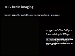
THG brain imaging
Depth scan through the prefrontal cortex of a mouse:
Image size 500 x 500 µm.Scanned depth 360 µm.Witte, S. Witte, A. Negrean, J.C. Lodder, C.P.J. de Kock, G.T. Silva, H.D. Mansvelder, and M.L. Groot, Label-free live brain imaging and targeted patching with third-harmonic generation microscopy. Proceedings of the National Academy of Sciences of the United States of America, 2011. 108(15): p. 5970-5975.