
Main Function of the digestive system Digestion the process by which your body breaks down the food you eat into substances that it can take absorb take in and use Organs of the Digestive system ID: 909083
Download Presentation The PPT/PDF document "The Digestive System Advanced Health" is the property of its rightful owner. Permission is granted to download and print the materials on this web site for personal, non-commercial use only, and to display it on your personal computer provided you do not modify the materials and that you retain all copyright notices contained in the materials. By downloading content from our website, you accept the terms of this agreement.


Slide1
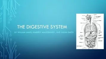
The Digestive System
Advanced Health
Slide2Main Function of the digestive system
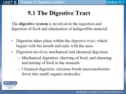
Digestion
- the process by which your body breaks down the food you eat into substances that it can take absorb (take in) and use.
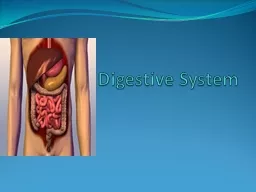
Slide3Organs of the Digestive system
Main Organs
Mouth (Oral Cavity)
Pharynx
Esophagus
Stomach
Small IntestineLarge Intestine (Colon)RectumAnus
Accessory Organs
Teeth and Tongue
Salivary Glands
Liver
Gallbladder
Pancreas
Appendix
Slide4Slide5Mouth (oral cavity)
The mouth is where food enters or is ingested into the digestive tract and the process of digestion begins.
The roof of the mouth is formed by the hard and soft palates and hanging down from the center of the soft palate is the uvula. The soft palate and the uvula, in most circumstances, prevent any food or liquid from entering the nasal cavities above the mouth.
The floor of the mouth consists of the tongue and its muscles.
The mouth also consists of the teeth (20 baby teeth and 32 permanent teeth, which are responsible for mastication (chewing) the food.
Slide6Salivary Glands
As we chew, the salivary glands around the mouth produce saliva that softens the food so it is easier to swallow.
Saliva also contains enzymes that start to break down and digest the food.
These glands are located outside of the digestive tube itself and must convey their secretions by ways of ducts into the track.
Slide7Swallowing
Swallowing occurs when you push some chewed food towards the back of your mouth.
As you swallow, two involuntary events occur:
The soft palate is pressed upwards to stop food from getting into your nose.
The epiglottis (flap of cartilage at the top of the windpipe), tilts down over the larynx to stop the food from entering the airways.
Slide8pharynx
Tube-like structure found in both the respiratory and digestive systems.
Because of its location behind the nasal cavities and mouth, it functions as part of the respiratory and digestive systems.
Air must pass through the pharynx on its way to the lungs and food must pass through on its way to the stomach.
Slide9Esophagus
Muscle-lined tube that connects the pharynx to the stomach.
The esophagus is about 9-10 inches (25 centimeters) long and less than an inch (2 centimeters) in diameter when relaxed.
Slide10Stomach
Muscle-walled pouch that food enters after it has been chewed, swallowed, and passed through the esophagus.
The sight, smell, and taste of food all start the production of gastric juices so that by the time the food reaches the stomach, it is ready to start digestion.
The gastric juices produced by the stomach contain acid and enzymes that break down proteins.
Once the food has been reduced to a liquid, the muscular ring at the exit of the stomach relaxes and the liquid enters the small intestine.
The mixture of churned food and gastric juices that leaves the stomach is called
chyme.
Stomach muscle contractions result in
peristalsis,
which propels food down the digestive tract.
Slide11The small intestine has three distinct regions:
1.
Duodenum
- mixes the chyme with enzymes and bile to break it down into molecules small enough to be absorbed
2.
Jejunum
- where food is absorbed
3.
Ileum
- absorbs vitamin B12
The part of the gastrointestinal tract between the stomach and the large intestine where most of the end absorption of food takes place.
Facts:
Roughly 7 meters (20 feet long)
Lined with protective mucus
Lining also consists of thousands of folds and tiny projections called villi
The primary function of the small intestine is the absorption of nutrients and minerals from food.
Once all of the useful molecules have been absorbed, the remaining liquid passes into the large intestine.
SMALL INTESTINE
Slide12Large intestine facts and anatomy
Facts:
The large intestine performs the vital functions of converting food into feces, absorbing essential vitamins produced by gut bacteria, and reclaiming water from feces.
About 1.5 meters (five feet long), but much larger diameter than small intestine.
It will process over 50 tons of consumed items over a lifetime.
Waste products can stay within the large intestine for two days or sometimes even more under certain circumstances.
Anatomy:
Slide13Large intestine
Chyme enters the large intestine from the small intestine.
Chyme passes through the cecum where it is mixed with beneficial bacteria that have colonized the large intestine throughout a person’s lifetime.
The chyme is then slowly moved through the four regions of the colon.
Most of the movement of chyme is achieved by slow waves of peristalsis over a period of several hours, but the colon can also be emptied quickly by stronger waves of mass peristalsis following a large meal.
While chyme moves through the large intestine, bacteria digest substances in the chyme that are not digestible by the human digestive system.
Bacterial fermentation converts the chyme into feces and releases vitamins including K, B1, B2, B6, B12, and biotin.
Vitamin K is almost exclusively produced by the gut bacteria and is essential in the proper clotting of blood. Gases such as carbon dioxide and methane are also produced as a byproduct of bacterial fermentation and lead to flatulence, or gas passed through the anus.
The absorption of water by the large intestine not only helps to condense and solidify feces, but also allows the body to retain water to be used in other metabolic processes.
Slide14The appendix
The appendix is a cul-de-sac of a tube connected to your cecum.
For years, many said the appendix had no purpose, but recent studies have found that the appendix may in fact act as a reservoir for good bacteria.
These bacteria contribute to keeping the gut healthy and helping you recover from infections.
Slide15The Rectum and anus
The rectum and anus are the final stages of the digestive tract.
The rectum is the last straight section of the large intestine before reaching the anus. The rectum is an 8-inch chamber that connects the colon to the anus. It is the rectum's job to receive stool from the colon, to let the person know that there is stool to be evacuated, and to hold the stool until evacuation happens. When anything (gas or stool) comes into the rectum, sensors send a message to the brain. The brain then decides if the rectal contents can be released or not.
The anus is the last part of the digestive tract. It is a 2-inch long canal surrounded by sphincter muscles that are important in allowing control of stool.
Slide16Slide17The Liver
The biggest internal organ is the liver.
Functions of the liver:
Breakdown of lipids and fat
Breaks down
Remove toxins
Recycles red blood cells and makes new plasma
Regulates blood glucose levels
Stores vitamins and minerals
Produces and secretes bile (a yellowish or greenish bitter liquid that helps dissolve fat as food is ingested in the intestines)
Slide18The GallBladder
A small hollow bag that lies behind the liver and stores the bile.
It is connected to the common bile duct by the cystic duct and the common bile duct connects the liver to the small intestine.
Bile is released into the small intestine when we eat and helps in the digestion of fats.
Bile contains a fatty material called cholesterol. If there is too much cholesterol in the bile, it may start to turn solid and form lumps called gallstones.
Some people with gallstones have their gall bladder removed (you do not need a gallbladder to live), but these people may have to go on a very low fat diet as they cannot digest fats properly.
Slide19The Pancreas
A large, carrot-shaped gland that lies just below your stomach.
Main function of the pancreas is to secrete insulin to break down sugars.
It also produces enzymes or digestive juices, which are secreted into the small intestine to further break down food after it has left the stomach.
The pancreas secretes the body’s own antacid, sodium bicarbonate to settle an upset stomach.
The head of the pancreas is on the right side of the abdomen and is connected to the duodenum (the first section of the small intestine) through a small tube called the pancreatic duct.
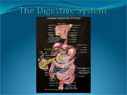
Slide20Label the Digestive System Diagram
1. Mouth (Oral Cavity)
2. Liver
3. Gallbladder
4. Large Intestine
5. Appendix
6. Anus7. Esophagus8. Stomach9. Pancreas10. Small Intestine11. Rectum
Slide21The Digestive Process
https://www.youtube.com/watch?v=08VyJOEcDos
Slide22What’s My Problem?
You are going to receive a worksheet.
Your job? Correctly identify15 common digestive system ailments.
Read the directions on the worksheet and begin!
We will check our work after.
Slide23Answers
1. Gastroesophageal Reflux Disease (GERD)
2. Nausea
3. Jaundice
4. Hepatitis
5. Flatulence
6. Diverticulitis7. Constipation8. Diarrhea
9. Colitis10. Cirrhosis11. Irritable Bowel Syndrome (IBS)12. Stomach Ulcer
13. Hemorrhoids
14. Anal Fissures
15. Hiatal Hernia