
Dr Ali Khazaal Jumaa FIBMS Internal Medicine FIBMS Clinical Hematology Gross anatomy and histology Bone marrow is a jellylike substance that fills the cavity left by the trabecular network of bone It accounts for about 4 5 of the total body weight of an individual It is resp ID: 998304
Download Presentation The PPT/PDF document "Bone marrow and hemopoiesis" is the property of its rightful owner. Permission is granted to download and print the materials on this web site for personal, non-commercial use only, and to display it on your personal computer provided you do not modify the materials and that you retain all copyright notices contained in the materials. By downloading content from our website, you accept the terms of this agreement.


1. Bone marrow and hemopoiesisDr. Ali Khazaal JumaaF.I.B.M.S (Internal Medicine)F.I.B.M.S (Clinical Hematology)
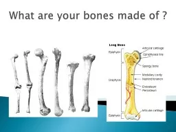
2. Gross anatomy and histologyBone marrow is a jelly-like substance that fills the cavity left by the trabecular network of bone. It accounts for about 4 – 5% of the total body weight of an individual. It is responsible for producing platelets, lymphocytes, erythrocytes, granulocytes and monocytes.Marrow has two principal functions; one is to produce blood cells and the other is to store fat. As a result, there are two types of marrow found in the body:1- The highly vascular red marrow which is hemopoietically active2- The fat rich yellow marrow that has significantly less hemopoietic centers and more adipocytes.
3. Red bone marrowClusters of hemopoietic cells known as hemopoietic islands are widely distributed throughout the loose connective tissue network observed in red marrow. These islands are found next to relatively large, thin walled, sinusoids that also communicate with nutrient vessels of the bone.Red marrow is most abundant in all skeletal structures from intrauterine life up until around the 5th year of life. As time progresses, red marrow is restricted to the central flat bones (i.e. cranial bones, clavicle, sternum, ribs, scapula, vertebrae, and pelvis) and the proximal ends of the proximal long bones of the upper and lower limbs.The supporting substance that supports the hemopoietic cells and adipocyte in the marrow is made up of reticulin. This is a fine collagen that is produced by mesenchyme derived reticular cells (fibroblast-like cells).
4.
5. Yellow bone marrowDepending on the age and hematological demand of an individual, the reticular cells become swollen as a result of increased lipid uptake. Subsequently, yellow marrow is formed. It contains mainly supportive connective tissue that provides scaffolding for the neurovascular structures that traverse the cavitation. There are also numerous adipocytes in addition to very few dormant hemopoietic clusters. These latent hemopoietic centers can be reactivated in the event of an increase demand for red blood cells.
6.
7. Blood supplyThe bone marrow is perfused by the same arteries that provide nutrients to the surrounding bone. A nutrient artery and two nutrient veins travel through nutrient canals to enter the bone marrow. The nutrient artery bifurcates after entering the marrow; each artery travels along the long axis of the bone in opposite directions. The artery subsequently branches out to supply the bony cortex. However, some of these thin-walled arterioles and their subsequent capillaries anastomose with venous sinus plexuses. The plexuses are tributaries to the collecting venules, which also drain to the central longitudinal vein. The central longitudinal vein then returns newly formed blood cells from the marrow pool to the systemic circulation.
8. InnervationThe nutrient canals – as well as both epiphyseal and metaphyseal foramina – also carry both unmyelinated and myelinated nerve fibers to the bone; and by extension, the marrow. Some of these fibers serve as vasa nervosa – small arteries that provide blood supply to peripheral nerves – as well as the hemopoietic tissue of the marrow.
9. Cell typesHistological analysis of the bone marrow will reveal an abundance of progenitor cells and their derivatives at different stages of development. Typically, the progenitor cells are larger than their end products. The suffix “-blast” is often used to denote that the cell line being referenced are the stem cells for that series (i.e. erythroblasts are the precursor cells for red blood cells [erythrocytes]).
10. BloodBlood is a bright to dark red, viscous, slightly alkaline (pH 7.4) fluid that accounts for approximately 7% of the total body weight. The total volume of blood of an average adult is about 5 L, and it circulates throughout the body within the confines of the circulatory system. Blood is a specialized connective tissue composed of cellular elements—red blood cells (RBCs; erythrocytes), white blood cells (WBCs; leukocytes), and platelets—suspended in a fluid component (the extracellular matrix), known as plasma.
11. PlasmaPlasma is a yellowish fluid in which cells, organic compounds, and electrolytes are suspended and/or dissolved. During coagulation, some of the clotting factors leave the plasma to become integrated into the clot. The remaining fluid, which no longer has the clotting factors is known as serum. The major component of plasma is water, constituting about 90% of its volume. Proteins compose 9%, and inorganic salts, ions, nitrogenous compounds, nutrients, and gases constitute the remaining 1%.
12. Plasma leaves the capillaries and small venules to enter the connective tissue spaces as extracellular fluid, which thus has a composition of electrolytes and small molecules similar to that in plasma. The concentration of proteins in extracellular fluid is much lower than that in plasma because it is difficult even for small proteins, such as albumin, to traverse the endothelial lining of a capillary or that of a venule. In fact, one of the proteins of plasma, albumin, is chiefly responsible for the establishment of blood’s colloid osmotic pressure, the force that maintains normal blood volume by opposing the movement of fluid from the capillaries and venules into the interstitial spaces.
13. Stem Cells, Progenitor Cells, and Precursor CellsThe least differentiated of the cells responsible for the formation of the formed elements of blood are stem cells; stem cells give rise to progenitor cells whose progeny are the precursor cells. All blood cells arise from pluripotent hemopoietic stem cells (PHSCs) (also known as hemopoietic stem cells [HSC]), which account for about 0.01% of the nucleated cell population of bone marrow. They undergo bursts of cell division, giving rise to more PHSCs as well as to two types of multipotent hemopoietic stem cells (MHSCs).
14. The two populations of MHSCs are colony-forming unit– lymphocyte (CFU-Ly), also known as common lymphoid progenitors, and CFU-GEMM (colony forming unit-granulocyte, erythrocyte, monocyte, megakaryocyte), also known as common myeloid progenitors. These two populations of MHSCs are responsible for the formation of a series of precursor cells, each of which gives rise to one specific type of the various types of blood cells.
15. Early stem cells may be recognized because they express the specific marker molecules CD34, CD59, CD133, and c-kit on their plasma membranes.Progenitor cells are unipotential (i.e., committed to forming a single cell line). Their mitotic activity and differentiation are controlled by specific hemopoietic factors. These cells have only limited capacity for self-renewal.Precursor cells arise from progenitor cells, are incapable of self-renewal, and have specific morphological characteristics that permit them to be recognized as the first cell of a particular cell line. Precursor cells undergo cell division and differentiation, eventually giving rise to a clone of mature cells.
16.
17. Hemopoietic Growth Factors (Colony-Stimulating Factors)Hemopoiesis is regulated by numerous growth factors produced by various cell types. Each factor acts on specific stem cells, progenitor cells, and precursor cells, generally inducing rapid mitosis, differentiation, or both. Most hemopoietic growth factors are glycoproteins.Three routes are used to deliver growth factors to their target cells: transport via the bloodstream (as endocrine hormones), secretion by stromal cells of the bone marrow near the hemopoietic cells (as paracrine hormones), direct cell–cell contact (as surface signaling molecules).
18. Site of OriginPrincipal ActionFactorsStromal cells of bone marrowStimulate proliferation of pluripotential and multipotential stem cellsStem cell factor (steelfactor, c-kit ligand)T cells; endothelial cellsPromotes CFU-GM mitosis and differentiationGM-CSFMacrophages; endothelial cellsPromotes CFU-G mitosis and differentiationG-CSFMonocytes; macrophages,endothelial cellspromotes proliferation ofPHSC, CFU-GEMM, and CFU-LyIL-1, IL-3, IL-6Activated T cellsStimulates activated T- and B-cell mitosisIL-2, IL-4T cells and NK cellsActivate B cells and monocytesγ-InterferonsKidney, LiverCFU-E differentiation; BFU-E mitosisErythropoietinLiver, Kidney, Bone marrowProliferation and differentiation of CFU-Meg and megakaryoblastsThrombopoietinCFU: colony forming unit (E: erythrocyte, G: granulocyte, Ly: lymphocyte, M: monocyte, Meg: megakaryocyte), CSF:colony-stimulating factor, BFU: Burst forming unit, IL: interleukin, NK: natural killer, PHSC: pluripotential hemopoietic stem cell.
19. ErythropoiesisErythrocytes (red blood cells), the smallest and most numerous cells of blood, have no nuclei and are responsible for the transport of oxygen and carbon dioxide to and from the tissues of the body.Each erythrocyte (RBC) resembles a biconcave shaped disk that is 7.5 μm in diameter, 2.0 μm thick at its widest region, and less than 1 μm thick at its center. This shape provides the cell with a large surface area relative to its volume, thus enhancing its capability for gaseous exchange. Although erythrocyte precursor cells within the bone marrow possess nuclei during development and maturation, the precursor cells or erythrocytes expel not only their nuclei but also all of their organelles prior to entering the circulation. Thus, mature circulating erythrocytes have no nuclei.
20. Erythropoiesis is under the control of several cytokines, namely steel factor, interleukin-3, and erythropoietin.Pathologically increased secretion of erythropoietin can cause secondary polycythemia, an increase in the total number of red blood cells in the blood, increasing its viscosity, reducing its flow rate, and thus impeding circulation.
21.
22.
23. GranulopoiesisGranulocytes are a category of white blood cells in the innate immune system characterized by the presence of specific granules in their cytoplasm. They are also called polymorphonuclear leukocytes (PMN, PML, or PMNL) because of the varying shape of the nucleus. The term polymorphonuclear leukocyte often refers specifically to neutrophil granulocytes, the most abundant of the granulocytes; the other types (eosinophils, basophils, and mast cells) have lower number of lobes.The proliferation and differentiation of myeloid progenitor cells are under the influence of G-CSF, IL-3, and GM-CSF
24. Types of granulopoiesisSteady state granulopoiesisSteady state granulopoiesis is a term used to describe the normal daily production of granulocytes. Granulocytes are short lived cells (their lifespan is between 6 and 8 hours) with a high cell turnover.Emergency granulopoiesisSteady state granulopoiesis is switched to a program termed emergency granulopoiesis after a major insult to the organism, usually a bacterial infection.
25. There are three types of granulocytes :NeutrophilsBasophilsEosinophils
26. NeutrophilsNeutrophils are the most abundant type of white blood cells, constituting 60% to 65%, and about 12–15 micrometres in diameter. Once neutrophils have received the appropriate signals, it takes them about thirty minutes to leave the blood and reach the site of an infection to play an important role in phagocytosis. Neutrophils do not return to the blood; they turn into pus cells and die. Mature neutrophils are smaller than monocytes, and have a segmented nucleus with several sections(two to five segments); each section is connected by chromatin filaments. Neutrophils do not normally exit the bone marrow until maturity, but during an infection neutrophil precursors called myelocytes and promyelocytes are released.
27. EosinophilsEosinophils have kidney-shaped lobed nuclei (two to four lobes). The number of granules in an eosinophil can vary because they have a tendency to degranulate while in the blood stream. Eosinophils play a crucial part in the killing of parasites (e.g., enteric nematodes). These cells also have a limited ability to participate in phagocytosis, they are professional antigen-presenting cells, they regulate other immune cell functions (e.g., CD4+ T cell, dendritic cell, B cell, mast cell, neutrophil, and basophil functions), they are involved in the destruction of tumor cells, and they promote the repair of damaged tissue.
28. BasophilsBasophils are one of the least abundant cells in bone marrow and blood (occurring at less than two percent of all cells). Like neutrophils and eosinophils, they have lobed nuclei; however, they have only two lobes, and the chromatin filaments that connect them are not very visible. Basophils have receptors that can bind to IgE, IgG, complement, and histamine. The cytoplasm of basophils contains a varied amount of granules; these granules are usually numerous enough to partially conceal the nucleus. Granule contents of basophils are abundant with histamine, heparin, platelet-activating factor, and other substances.
29. When an infection occurs, mature basophils will be released from the bone marrow and travel to the site of infection. When basophils are injured, they will release histamine, which contributes to the inflammatory response that helps fight invading organisms. Histamine causes dilation and increased permeability of capillaries close to the basophil. Injured basophils and other leukocytes will release another substance called prostaglandins that contributes to an increased blood flow to the site of infection. Both of these mechanisms allow blood-clotting elements to be delivered to the infected area (this begins the recovery process and blocks the travel of microbes to other parts of the body). Increased permeability of the inflamed tissue also allows for more phagocyte migration to the site of infection so that they can consume microbes.
30. MonocytesMonocytes are a type of leukocyte, or white blood cell. They are the largest type of leukocyte. Monocytes are produced by the bone marrow fromprecursors called monoblasts. Monocytes circulate in the bloodstream for about one to three days andthen typically move into tissues throughout the body where they differentiate into macrophages monocyte-derived dendritic cells, osteoclasts, and microglia. They constitute between three and eight percent of the leukocytes in the blood. They have a role in phagocytosis, antigen presentation and cytokine production.
31. Platelet FormationThe formation of platelets is under the control of thrombopoietin,which induces the development and proliferation of giant cells known as megakaryoblasts.
32. Platelets They are the smallest of the three major types of blood cells.Platelets are only about 20% of the diameter of red blood cells.Platelet ProductionPlatelets are produced in the bone marrow, the same as the red cells and most of the white blood cells. Platelets are produced from very large bone marrow cells called megakaryocytes. As megakaryocytes develop into giant cells, they undergo a process of fragmentation that results in the release of several thousand platelets per megakaryocyte. The remaining cytoplasm and nucleus of the megakaryocyte degenerate and are phagocytosed by macrophages.The dominant hormone controlling megakaryocyte development is thrombopoietin (often abbreviated as TPO).
33. Platelet StructurePlatelets are actually not true cells but merely circulating fragments of cells. But even though platelets are merely cell fragments, they contain many structures that are critical to stop bleeding. They contain proteins on their surface that allow them to stick to breaks in the blood vessel wall and also to stick to each other. They contain granules that can secrete other proteins required for creating a firm plug to seal blood vessel breaks. When platelets are stimulated by a break in the blood vessel wall they change shape. They become round and extend long filaments to make contact with the broken blood vessel wall or with other platelets. With these long filaments, platelets then form a plug to seal the broken blood vessel.
34. LymphocytesA lymphocyte is a type of white blood cell in the immune system. They include:natural killer cells (which function in cell-mediated, cytotoxic innate immunity), T cells (for cell-mediated, cytotoxic adaptive immunity), and B cells (for humoral, antibody-driven adaptive immunity).They are the main type of cell found in lymph, which prompted the name "lymphocyte".Lymphocytes make up between 18% and 42% of circulating leukocytes.Lymphocytes are indistinguishable under the microscope, but by immunophenotyping.
35. FunctionsT cells (thymus cells) and B cells (bone marrow cells) are the major cellular components of the adaptive immune response. T cells are involved in cell-mediated immunity, whereas B cells are primarily responsible for humoral immunity (relating to antibodies). The function of T cells and B cells is to recognize specific "non-self" antigens, during a process known as antigen presentation. Once they have identified an invader, the cells generate specific responses to eliminate specific pathogens or pathogen-infected cells. B cells respond to pathogens by producing large quantities of antibodies (after maturation into plasma cells) which then neutralize foreign objects like bacteria and viruses.
36. In response to pathogens some T cells, called T helper cells, produce cytokines that direct the immune response, while other T cells, called cytotoxic T cells, produce toxic granules that contain powerful enzymes which induce the death of pathogen-infected cells. Following activation, B cells and T cells leave a lasting legacy of the antigens they have encountered, in the form of memory cells. Throughout the lifetime of an animal, these memory cells will "remember" each specific pathogen encountered, and are able to mount a strong and rapid response if the same pathogen is detected again; this is known as acquired immunity.
37. LymphocytopoiesisThe multipotential stem cell CFU-Ly divides in the bone marrow to form the three unipotential progenitor cells, CFU-LyB, CFU-LyT, and CFU-NK cells.CFU-LyB divides several times, giving rise to immunocompetent B lymphocytes expressing specific surface markers, like CD19 and CD20. The process of B cell maturation is partially controlled by IL-7 and Pax5 transcription factor.CFU-LyT cells undergo mitosis, forming immunoincompetent T cells, which travel to the cortex of the thymus, where they proliferate, mature, and begin to express specific cell surface markers, like CD3. The process of T cell maturation is partially controlled by IL-7 and GATA3 transcription factor.CFU-NK cells also migrate to a specific region in the bone marrow where they will become immunocompetent. The process of NK cell maturation is partially controlled by IL-12 and IL-15.
38.