
Overall annual incidence of CVST of 0213 per 100000 people 2 CVST prevalence of 292 among 140 cases with a skull fracture that crossed a cerebral venous sinus 2 11 Higher incidence among females and neonates ID: 913739
Download Presentation The PPT/PDF document "The dural or cerebral venous sinuses a..." is the property of its rightful owner. Permission is granted to download and print the materials on this web site for personal, non-commercial use only, and to display it on your personal computer provided you do not modify the materials and that you retain all copyright notices contained in the materials. By downloading content from our website, you accept the terms of this agreement.


Slide1
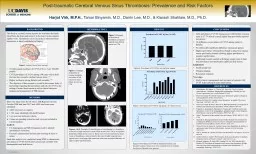
The dural or cerebral venous sinuses are structures that drain blood from the brain and return it to the heart via the internal jugular veins. Thrombosis can occur in these structures from hemostasis or direct endothelial cell injuryOverall annual incidence of CVST of 0.2-1.3 per 100,000 people.2CVST prevalence of 29.2% among 140 cases with a skull fracture that crossed a cerebral venous sinus.2, 11Higher incidence among females and neonates.2-10The objective of this research is to add to the current body of knowledge regarding the prevalence and risk of CVST in the setting of acute, blunt trauma to aid in clinical decision-making and management of TBI patients.
BACKGROUND
METHODS
Figure 2
. Example of thrombosis identified in superior sagittal sinus (black arrow) on CTA.
METHODS (CONT.)
RESULTS
CONCLUSIONS
Kristoffersen, ES, Harper, C., Vetvik, KG, Faiz, KW. (2018). Cerebral venetrombose – forekomst, diagnostikk og behandling. Tidsskrift for Den Norske Legeforening. doi: 10.4045/tidsskr.17.1047Ferro JM, Canhao P. Cerebral venous sinus thrombosis: update on diagnosis and management. Stroke. 2014;16(523). Bousser M, Ferro JM. Cerebral venous thrombosis: an update. The Lancet Neurology. 2007;6(2): 162-170.Ferro JM, Canhao P, Stam J, Bousser M, Barinagarrementeria F. Prognosis of cerebral vein and dural sinus thrombosis. Stroke. 2004;35: 664-670. Filippidis A, Kapsalaki E, Patramani G, Fountas KN. Cerebral venous sinus thrombosis: review of the demographics, pathophysiology, current diagnosis, and treatment. Journal of Neurosurgery. 2009;27(5): E3. Stam J. Thrombosis of the cerebral veins and sinuses. NEJM. 2005;352: 1791-1798.Saposnik G, Barinagarrementeria F, Brown RD, Bushnell CD, Cucchiara B, Cushman M, deVeber G, Ferro JM, Tsai FY. Diagnosis and management of cerebral venous thrombosis. Stroke. 2011;42: 1158-1192.Wasay M, Dai AI, Ansari M. Cerebral venous sinus thrombosis in children: a multicenter cohort from the United States. Journal of Child Neurology. 2007;23(1).Moll S, Waldron B. Cerebral and sinus vein thrombosis. Circulation. 2014;130: e68-e70.Stiefel D, Eich G, Sacher P. Posttraumatic dural sinus thrombosis in children. European Journal of Pediatric Surgery. 2000;10(1): 41-44.Delgado Almandoz JE, Kelly HR, Schaefer PW, Lev MH, Gonzalez RG, Romero JM. Prevalence of traumatic dural venous sinus thrombosis in high-risk acute blunt head trauma patients evaluated with multidetector CT venography. Radiology. 2010;255(2).
REFERENCES
Data were taken from the UC Davis TBI Registry between October 2008 and June 2017 and 4,886 total cases were identified.4,886 total cases in registry1,748 cases identified with skull fractures144 cases met inclusion criteria9 cases are pending evaluation and were not included in current analysisAnalysisCT Angiograms and MR Venograms used to identify intraluminal occlusionsFractures characterized by bone and crossing of sinus or sutureFinal data analysis was conducted using SPSS to characterize prevalence of CVST by fracture location and correlate with demographics and risk factors.
Harjot Virk, M.P.H.,
Tamar Binyamin, M.D., Darrin Lee, M.D., &
Kiarash
Shahlaie
, M.D., Ph.D.
Post-traumatic Cerebral Venous Sinus Thrombosis: Prevalence and Risk Factors
Total prevalence of CVST among cases with fracture crossing sinus is 57.7% which is much higher than previously reported prevalence.
No difference in prevalence of CVST among males vs. females.
No statistically significant difference among age groups.
Highest prevalence of thrombosis found in transverse sinuses versus previously research showing higher prevalence in superior sagittal sinus.
Additional research needed with larger sample sizes to find true prevalence and statistically significant risk factors.
Limitations
Small sample size
Missing imaging
Researcher expertise
Next steps
Study clinical management and outcomes of patients with CVST in the setting of acute head trauma.
Figure 3
. Example of thrombosis causing complete occlusion of left transverse sinus (black arrows) on CTA.
Figures 4 & 5
. Example of identification of extraluminal vs. thrombotic occlusion of right transverse sinus (white arrows) on CTA by an epidural hematoma (black arrows) on CT. These cases were not included in final count of thromboses. Also pictured is fracture of right temporal bone (green arrow) overlying right transverse sinus and associated diastasis of right
occipito
-mastoid suture.
Figure 1
. Cerebral Venous Sinus Anatomy
1
.
Figure 6. Prevalence of CVST by sex and total sample
.
Figure 7. Prevalence of CVST by age group
. “X” denotes 0 cases.
Figure 8. Prevalence of CVST by thrombus location and fracture location. Some cases were counted twice if multiple fractures or thrombi were present.