
Whole skeleton and bones associated with the Skull Copyright c The McGrawHill Companies Inc Permission required for reproduction or display 8 2 Unit 3 Bones chapters 8 In Chapter 8 you will learn ID: 775250
Download Presentation The PPT/PDF document " Bones Bones Bones Chapter 8" is the property of its rightful owner. Permission is granted to download and print the materials on this web site for personal, non-commercial use only, and to display it on your personal computer provided you do not modify the materials and that you retain all copyright notices contained in the materials. By downloading content from our website, you accept the terms of this agreement.


Slide1
Bones BonesBones
Slide2Chapter 8Whole skeleton and bones associated with the Skull
Copyright (c) The McGraw-Hill Companies, Inc. Permission required for reproduction or display.
8-2
Slide3Unit 3 Bones (chapters 8 )
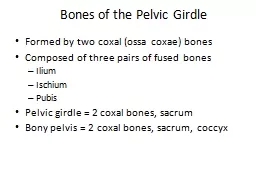
In Chapter 8 you will learn:Bones of the Adult Skeletal System a. Skull bones slides 1-20 b. Vertebral and Thoracic Bones c. Bones of the Pectoral and Upper Limb d. Bones of the Pelvic Girdle and Lower Limb2. Surface Features of Bones
7-
3
Slide48-4
The Skeletal System
overview of the skeletonthe skull the vertebral column and thoracic cagethe pectoral girdle and upper limbthe pelvic girdle and lower limb
Figure 8.1a
Copyright © The McGraw-Hill Companies, Inc. Permission required for reproduction or display.
Skull
Frontal bone
Pectoral
girdle
Clavicle
Maxilla
Parietal bone
Mandible
Mandible
Humerus
Femur
Fibula
Ulna
Radius
Scapula
Hip bone
Sacrum
Patella
Carpus
Thoracic
cage
Pelvis
Sternum
Ribs
Costal cartilages
Phalanges
Metatarsal bones
Phalanges
Coccyx
Vertebral column
Metacarpal
bones
Tarsus
(a) Anterior view
Tibia
Slide58-5
Overview of the Skeleton
two regions of the skeleton
axial skeleton
– forms the central supporting axis of the body
skull, auditory ossicles, hyoid bone, vertebral column, and thoracic cage (ribs and sternum)
appendicular skeleton
– includes the bones of the upper limb and pectoral girdle, and the bones of the lower limb and pelvic girdle
number of bones
206
in typical adult skeleton
varies with development of sesamoid bones (patella)
bones that form within some tendons in response to stress
varies with presence of sutural (wormian) bones in skull
extra bones that develop in skull suture lines
270
bones at birth, decreases with fusion
surface markings defined in Table 8.2
ridges, spines, bumps, depressions, canals, pores, slits, cavities, and articular surfaces
Slide68-6
Axial and Appendicular Skeleton
axial skeleton is colored tanskull, vertebrae, sternum, ribs, sacrum and hyoidappendicular skeleton is colored greenpectoral girdleupper extremitypelvic girdlelower extremity
Copyright © The McGraw-Hill Companies, Inc. Permission required for reproduction or display.
Skull
Frontal bone
Clavicle
Maxilla
Parietal bone
Mandible
Occipital bone
Mandible
Humerus
Femur
Fibula
Ulna
Radius
Scapula
Clavicle
Scapula
Hip bone
Sacrum
Patella
Carpus
Pelvis
Sternum
Ribs
Costal cartilages
Phalanges
Metatarsal bones
Phalanges
Coccyx
(b) Posterior view
Pectoral
girdle
Thoracic
cage
Vertebral column
Metacarpal
bones
Tarsus
(a) Anterior view
Tibia
Figure 8.1
Slide78-7
Anatomical Features of Bones
Copyright © The McGraw-Hill Companies, Inc. Permission required for reproduction or display.
Sinuses
Crest
Foramen
Foramen
Fossae
Head
Tubercle
Crest
Tuberosity
Line
Head
Fovea
Trochanters
Fossae
(a) Skull (lateral view)
Epicondyles
Condyles
Alveolus
Spine
Condyle
Process
Lines
Meatus
(b) Scapula (posterior view)
(c) Femur
(posterior view)
(d) Humerus (anterior view)
Process
Spine
Figure 8.2
Slide88-8
The Skull
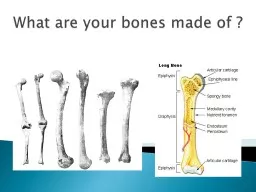
skull – the most complex part of the skeleton
22 bones joined together by sutures (immovable joints)
8 cranial bones surround cranial cavity which encloses the brain
other cavities – orbits, nasal cavity, oral (buccal) cavity, middle-, and inner ear cavities, and paranasal sinuses
paranasal sinuses – frontal, sphenoid, ethmoid, and maxillary
lined by mucous membrane and air-filled
lighten the anterior portion of the skull
act as chambers that add resonance to the voice
foramina – holes that allow passage for nerves and blood vessels
14 facial bones support teeth, facial and jaw muscles
Slide98-9
Major Skull Cavities
Figure 8.7
Copyright © The McGraw-Hill Companies, Inc. Permission required for reproduction or display.
Frontal bone
Ethmoid bone
Middle
Superior
Inferior
Maxilla
Nasal cavity
Mandible
Vomer
Orbit
Cranial cavity
Nasal
conchae
Oral
cavity
Maxillary
sinus
Zygomatic
bone
Ethmoid
air cells
Slide108-10
Cranial Fossa
cranium (braincase) – protects the brain and associated sense organsswelling of the brain inside the rigid cranium may force tissue through foramen magnum resulting in death consists of two parts: the calvaria (skullcap) and the cranial basebase is divided into three basins that comprise the cranial flooranterior cranial fossa holds the frontal lobe of the brainmiddle cranial fossa holds the temporal lobes of the brainposterior cranial fossa contains the cerebellum
Copyright © The McGraw-Hill Companies, Inc. Permission required for reproduction or display.
(a) Superior view
Posterior cranial fossa
Middle cranial fossa
Anterior cranial fossa
Cerebellum
Temporal lobe
Frontal lobe
(b) Lateral view
Anterior cranial
fossa
Middle cranial
fossa
Posterior cranial
fossa
Figure 8.9
Slide118-11
Frontal Bone
forms forehead and part of the roof of the craniumcoronal suture – posterior boundary of frontal bonesupraorbital margin forms roof of the orbitsupraorbital foramen provides passage for nerve, artery, and veinglabella – smooth area above root of the nosecontains frontal sinus
Figure 8.3
Copyright © The McGraw-Hill Companies, Inc. Permission required for reproduction or display.
Frontal bone
Coronal suture
Lacrimal bone
Squamous suture
Infraorbital foramen
Vomer
Mandible
Sphenoid bone
Ethmoid bone
Nasal bone
Zygomatic bone
Maxilla
Mental foramen
Temporal bone
Glabella
Mental protuberance
Parietal bone
Middle nasal
concha
Inferior nasal
concha
Supraorbital
margin
Supraorbital
foramen
Slide128-12
Parietal Bone
form most of cranial roof and part of its lateral wallsbordered by 4 suturessagittal – between parietal bonescoronal – at anterior marginlambdoid – at posterior marginsquamous – at lateral bordertwo temporal lines serve as attachment of the temporalis muscle
Figure 8.4a
Copyright © The McGraw-Hill Companies, Inc. Permission required for reproduction or display.
Parietal bone
Frontal bone
Lambdoid suture
Sphenoid bone
Temporal bone
Zygomatic process
External acoustic meatus
Mandible
Temporal lines
Coronal suture
Ethmoid bone
Lacrimal bone
Nasal bone
Infraorbital foramen
Zygomatic bone
Temporal process
Zygomaticofacial foramen
Maxilla
Mental foramen
Occipital bone
Squamous suture
Mandibular condyle
Styloid process
Mastoid process
(a) Right lateral view
Copyright © The McGraw-Hill Companies, Inc. Permission required for reproduction or display.
Frontal bone
Coronal suture
Sagittal suture
Sutural bone
Parietal bone
Parietal foramen
Lambdoid suture
Occipital bone
Posterior
Anterior
Figure 8.6
Slide138-13
Temporal Bone
lateral wall and part of floor of cranial cavitysquamous partencircled by squamous suturezygomatic processmandibular fossa tympanic partexternal auditory meatusstyloid processmastoid partmastoid processmastoiditis from ear infectionmastoid notchstylomastoid foramenmastoid foramen
Figure 8.4a
Copyright © The McGraw-Hill Companies, Inc. Permission required for reproduction or display.
Parietal bone
Frontal bone
Lambdoid suture
Sphenoid bone
Temporal bone
Zygomatic process
External acoustic meatus
Mandible
Temporal lines
Coronal suture
Ethmoid bone
Lacrimal bone
Nasal bone
Infraorbital foramen
Zygomatic bone
Temporal process
Zygomaticofacial foramen
Maxilla
Mental foramen
Occipital bone
Squamous suture
Mandibular condyle
Styloid process
Mastoid process
(a) Right lateral view
Slide148-14
Petrous Portion of Temporal Bone
petrous partpart of cranial floorseparates middle from posterior cranial fossahouses middle and inner ear cavitiesreceptors for hearing and sense of balanceinternal auditory meatus - opening for CN VII (vestibulocochlear nerve)carotid canaljugular foramen
Copyright © The McGraw-Hill Companies, Inc. Permission required for reproduction or display.
(b) Superior view of cranial floor
Cribriform foramina
Crista galli
Diploe (spongy bone)
Sphenoid bone
Temporal bone
Parietal bone
Occipital bone
Sella turcica
Frontal bone
Optic foramen
Foramen rotundum
Foramen ovale
Foramen spinosum
Foramen magnum
Jugular foramen
Hypoglossal canal
Internal acoustic
meatus
Groove for
venous sinus
Petrous part of
temporal bone
Cribriform plate
of ethmoid bone
Figure 8.5b
Slide158-15
Right Temporal Bone
Mastoid part
Mastoid process
(a) Lateral surface
(b) Medial surface
Styloid process
Squamous suture
Styloid process
Tympanic part
Squamous part
Squamous suture
Zygomatic process
Mandibular fossa
Mastoid process
Squamous part
Petrous part
Mastoid notch
External acoustic
meatus
Zygomatic
process
Copyright © The McGraw-Hill Companies, Inc. Permission required for reproduction or display.
Internal acoustic
meatus
Figure 8.10
Slide168-16
Occipital Bone
rear and base of skullforamen magnum holds spinal cordbasilar partskull rests on atlas at occipital condyleshypoglossal canal transmits hypoglossal nerve (CN XII) supplying tongue musclescondylar canalexternal occipital protuberance for nuchal ligamentsuperior and inferior nuchal lines mark neck muscles
Figure 8.5a
Copyright © The McGraw-Hill Companies, Inc. Permission required for reproduction or display.
Zygomatic bone
Lambdoid suture
Foramen magnum
Mastoid foramen
Jugular foramen
Stylomastoid foramen
Carotid canal
Intermaxillary suture
Palatine bone
Greater palatine foramen
Medial pterygoid plate
Lateral pterygoid plate
Foramen ovale
Foramen spinosum
Foramen lacerum
Incisive foramen
Sphenoid bone
Zygomatic arch
Mandibular fossa
Styloid process
External acoustic meatus
Mastoid process
Mastoid notch
Condylar canal
Temporal bone
Superior nuchal line
Inferior nuchal line
Occipital bone
(a) Inferior view
Parietal bone
Occipital condyle
Vomer
Posterior nasal
aperture
Palatine process
of maxilla
Basilar part of
occipital bone
External occipital
protuberance
Slide17Sphenoid Bone
bodygreater winglesser wingoptic foramenanterior clinoid processessuperior orbital fissure
8-17
Figure 8.5b
Copyright © The McGraw-Hill Companies, Inc. Permission required for reproduction or display.
Lesser wing
Greater wing
Foramen ovale
Body
Lateral pterygoid plate
Medial pterygoid plate
Pterygoid processes
(b) Posterior view
Dorsum sellae
Foramen
rotundum
Superior orbital
fissure
Copyright © The McGraw-Hill Companies, Inc. Permission required for reproduction or display.
(b) Superior view of cranial floor
Cribriform foramina
Crista galli
Diploe (spongy bone)
Sphenoid bone
Temporal bone
Parietal bone
Occipital bone
Sella turcica
Frontal bone
Optic foramen
Foramen rotundum
Foramen ovale
Foramen spinosum
Foramen magnum
Jugular foramen
Hypoglossal canal
Internal acoustic
meatus
Groove for
venous sinus
Petrous part of
temporal bone
Cribriform plate
of ethmoid bone
Figure 8.11b
Slide188-18
Sphenoid Bone
Figure 8.11a
Copyright © The McGraw-Hill Companies, Inc. Permission required for reproduction or display.
Optic foramen
Lesser wing
Greater wing
Sella turcica
Dorsum sellae
Foramen rotundum
Hypophyseal fossa
Foramen ovale
Foramen spinosum
(a) Superior view
Anterior clinoid
process
Copyright © The McGraw-Hill Companies, Inc. Permission required for reproduction or display.
(b) Superior view of cranial floor
Cribriform foramina
Crista galli
Diploe (spongy bone)
Sphenoid bone
Temporal bone
Parietal bone
Occipital bone
Sella turcica
Frontal bone
Optic foramen
Foramen rotundum
Foramen ovale
Foramen spinosum
Foramen magnum
Jugular foramen
Hypoglossal canal
Internal acoustic
meatus
Groove for
venous sinus
Petrous part of
temporal bone
Cribriform plate
of ethmoid bone
Figure 8.5b
foramen rotundum
foramen ovale
foramen lacerum
posterior nasal apertures
or
choanae
medial pterygoid plate
lateral pterygoid plate
sphenoid sinus
Slide198-19
Sphenoid Bone
sphenoid sinus
Figure 8.4b
Copyright © The McGraw-Hill Companies, Inc. Permission required for reproduction or display.
Coronal suture
Frontal bone
Sphenoid sinus
Frontal sinus
Crista galli
Parietal bone
Temporal bone
Internal acoustic meatus
Jugular foramen
Hypoglossal canal
Mandibular foramen
Styloid process
Squamous suture
Lambdoid suture
Sella turcica
Occipital bone
Vomer
Palatine bone
Maxilla
Mandible
Mental spines
Nasal bone
(b) Median section
Cribriform plate of
ethmoid bone
Perpendicular plate
of ethmoid bone
Palatine process
of maxilla
Figure 8.5a
Copyright © The McGraw-Hill Companies, Inc. Permission required for reproduction or display.
Zygomatic bone
Lambdoid suture
Foramen magnum
Mastoid foramen
Jugular foramen
Stylomastoid foramen
Carotid canal
Intermaxillary suture
Palatine bone
Greater palatine foramen
Medial pterygoid plate
Lateral pterygoid plate
Foramen ovale
Foramen spinosum
Foramen lacerum
Incisive foramen
Sphenoid bone
Zygomatic arch
Mandibular fossa
Styloid process
External acoustic meatus
Mastoid process
Mastoid notch
Condylar canal
Temporal bone
Superior nuchal line
Inferior nuchal line
Occipital bone
(a) Inferior view
Parietal bone
Occipital condyle
Vomer
Posterior nasal
aperture
Palatine process
of maxilla
Basilar part of
occipital bone
External occipital
protuberance
Slide208-20
Ethmoid Bone
Figure 8.12
Supraorbital foramen
Orbital plate of ethmoid bone
Lacrimal bone
Optic foramen
Orbital plate of frontal bone
Lesser wing of sphenoid bone
Frontal process of maxilla
Superior orbital fissure
Roof of
orbit
Orbital process of
palatine bone
Orbital surface of
maxilla
Floor of
orbit
Medial
wall
Zygomatic process
of frontal bone
Greater wing of
sphenoid bone
Orbital surface of
zygomatic bone
Inferior orbital
fissure
Lateral wall
of orbit
Infraorbital
foramen
Copyright © The McGraw-Hill Companies, Inc. Permission required for reproduction or display.
between the eyes
contributes to medial wall of orbit
lateral walls and roof of nasal cavity, and nasal septum
three major portions
of this porous, delicate bone
perpendicular plate
forms superior two-thirds of nasal septum
cribriform plate
– forms roof of nasal cavity
crista galli
– attachment point for meninges
cribriform (olfactory) foramina
labyrinth –
large mass on each side of perpendicular plate
ethmoid cells
in the make up ethmoid sinuses
orbital plate
Figure 8.14
Orbital plate
Crista galli
Cribriform
plate
Cribriform
foramina
Ethmoidal
cells
Perpendicular
plate
Superior
nasal concha
Middle
nasal concha
Copyright © The McGraw-Hill Companies, Inc. Permission required for reproduction or display.
Slide218-21
Ethmoid Bone
superior and middle concha
perpendicular plate of nasal septum
Copyright © The McGraw-Hill Companies, Inc. Permission required for reproduction or display.
Coronal suture
Frontal bone
Sphenoid sinus
Frontal sinus
Crista galli
Parietal bone
Temporal bone
Internal acoustic meatus
Jugular foramen
Hypoglossal canal
Mandibular foramen
Styloid process
Squamous suture
Lambdoid suture
Sella turcica
Occipital bone
Vomer
Palatine bone
Maxilla
Mandible
Mental spines
Nasal bone
(b) Median section
Cribriform plate of
ethmoid bone
Perpendicular plate
of ethmoid bone
Palatine process
of maxilla
Frontal sinus
Inferior
Anterior nasal spine
Maxilla
Incisive foramen
Lacrimal bone
Nasal bone
Nasal cartilages
Superior
Middle
Sphenoid sinus
Palatine bone
Sphenoid bone
Crista galli
Sella turcica
Cribriform plate
Cribriform foramina
Occipital bone
Lip
Frontal bone
Incisor
Nasal conchae:
Copyright © The McGraw-Hill Companies, Inc. Permission required for reproduction or display.
Figure 8.4b
Figure 8.13