

Module Three Anatomy and Physiology Holdorf PhD MPA RDMS ObGyn Ab BR RVT LRTAS CCP Module Three Anatomy and Physiology Anatomic Layers From Anterior to Posterior Skin Subcutaneous premammary layer ID: 774838
Download Presentation The PPT/PDF document " Sonography of the Breast Part II" is the property of its rightful owner. Permission is granted to download and print the materials on this web site for personal, non-commercial use only, and to display it on your personal computer provided you do not modify the materials and that you retain all copyright notices contained in the materials. By downloading content from our website, you accept the terms of this agreement.


Slide1
Sonography of the Breast Part II
Module Three
Anatomy and Physiology
Holdorf
PhD, MPA, RDMS (Ob/Gyn, Ab, BR), RVT, LRT(AS,) CCP
Slide2Module Three: Anatomy and Physiology
Slide3Slide4Anatomic Layers
From Anterior to Posterior
Skin
Subcutaneous (premammary) layer
Mammary Layer
Retromammary Space
Muscle Layers (pectoralis Major m. and Pectoralis minor m.)
Chest wall (ribs and intercostal muscles)
Slide5Skin
The skin is composed of the epidermis and dermis layers
The thickness is 0.5 to 2 mm
It is slightly thicker in young females and thins with age.
NIPPLE
Consists of dense connective tissue and erectile muscle.
It contains many sensory nerve endings.
15 to 20 collecting (lactiferous) duct openings may be seen (each of which arise from a breast lobe)
AREOLA
Circular area of dark pigmentation seen around the nipple.
Consists of smooth muscle.
Slightly thicker than surrounding skin.
Contains Montgomery glands- sebaceous glands seen as small bumps in the areola.
Slide61. Subcutaneous (Premammary Layer)
2. Superficial layer
3. Deep layer
4. Superficial fascia
5. Mammary Layer
Slide7Layers of the breast
Slide8Subcutaneous (Premammary ) Layer
Lies just beneath the skin extending to the mammary layer
Consists primarily of fat
It is not seen posterior to the nipple
Amount of fat increases with age, pregnancy, and obesity
Cooper’s ligaments appear as prominent structures within the subcutaneous layer.
Slide9Superficial Fascia
The breast tissue is completely contained between the layers of the superficial fascia.
At the breast, the superficial fascia divides into the superficial and deep layers
The superficial layer of the superficial fascia is simply known as the superficial fascia.
The superficial fascia is contained within the subcutaneous layer anterior to the mammary layer.
Slide10Mammary Layer
The mammary layer is also known as the parenchymal or Glandular layer.
A portion of glandular tissue extends into the axilla. This is known as the axillary Tail of Spence.
The mammary layer is composed of two types of tissue:
Stroma- is the supportive tissue of the breast
Consists of interlobular fat and connective tissue (Cooper’s ligaments, loose and dense connective tissue)
Epithelium – is the functional tissue
Consist of acini, lobules, TDLU’s, lobes and lactiferous ducts.
Slide11Mammary layer- continued
Cooper’s Ligaments (Suspensory Ligaments)-
Part of the stoma and supportive tissue of he Mammary layer
Provide the architectural “framework” of the breast
Run between the superficial and deep layers of the superficial fascia.
Acini
Also called Acinus or Acinar cell
Smallest functional unit of the breast
Milk-producing gland
Hundreds of acini in each breast
Each acini gives rise to a ductule or terminal duct
Slide12Lobule
Is composed of approximately 30 acini, intralobular terminal ducts, and intralobular stromal tissue (loose connective tissue).
TDLU
Terminal Duct Lobular Unit
TLDU is made up of: Lobule, intralobular terminal ducts, extralobular terminal duct
Usually measures 2.0 mm or less
Nearly all breast pathology originates in the TDLU
.
Lobe
Several lobules (TDLUs) make up a breast lobe
15 to 20 lobes in each breast
One lactiferous duct emerges from each lobe and travels toward the nipple.
Slide13Mammary Layer (continued)
Lactiferous Ducts-
Transport milk from the acini to the nipple
Begin with the Ductule of Terminal Duct which arises from the acinar cell
Intralobular terminal Duct-within the lobule
Extralobular Terminal Duct- outside the lobule
Interlobular ducts travel between the lobes
The duct enlarges slightly beneath the areola forming the Lactiferous Sinus.
Collecting Duct empties milk from the nipple.
Slide14Layers of the breast
Slide15Cross section of Breast Duct lumen
Slide16Lactiferous ducts are lined with a double layer of epithelial cells
Epithelium (inner layer)
Myoepithelium
The epithelial cells are supported by a basement membrane (adventitia) which is the outer fibrous portion of the duct
The function of the my0epithelium is to produce milk within the ducts toward the nipple.
Deep Fascia
The deep layer of the superficial fascia is often referred to as the deep fascia.
Located within the retromammary space posterior to the mammary layer.
Maintaining integrity of the deep fascia is important in deterring the spread of cancer to the chest wall.
Slide17Retromammary Space
Space between the posterior margin of the mammary layer and the pectoral muscles
Contains thin layer of fat
Amount of fat increases with age, pregnancy, and obesity.
Also contains the deep layer of the superficial fascia.
This layer allows movement of the breast over the chest wall.
Slide18Muscles
Pectoralis Major arises from the clavicle and costal cartilage of the sternum, attaching to the proximal humerus.
Pectoralis Minor arises from the 3
rd
, 4
th
, and 5
th
ribs attaching to the scapula.
The pectoral (muscular) fascia encloses the chest muscles and may appear deep to the retromammary layer.
Pectoralis Major muscle is located anterior to pectoralis minor muscle. Both are found immediately posterior to the breast tissue.
Slide19Chest Wall
Ribs are located posterior to the pectoral muscles.
In small breasted females, it is important not to confuse a rib with an intra-mammary tumor on a physical or sonographic examination.
Intercostal muscles are located between the ribs.
Deep to the chest wall layer is the lung.
Slide20Slide21Deltoid, Pectoralis Major M.
Slide22Slide23Slide24Standard Anatomic Reference
Quadrant Method
Each breast can be divided into quadrants (4):
UO – Upper Outer
UI – Upper Inner
LO- Lower Outer
LI – Lower Inner
Glandular tissue is usually thicker in the Upper-Outer quadrant of both breasts
Therefore, a larger percentage of cancers are found there.
Slide25Clock method
Regions of the breast are correlated with positions of a clock. This method allows a more precise location to be documented.
Correlating clock locations from right to left side is important in evaluating the breast for symmetry. For example, the 10:00 position in the right breast correlates with the 2:00 position on the left.
Slide26Slide27Slide28Embryologic Development
The Early mammary gland begins development during the 4
th
week of embryologic life. The glandular tissue of the breast begins to evolve into epithelial buds that eventually form approximately 20 epithelial cords (lobes). At 15 weeks gestation, testosterone in the male fetus prohibits further breast development and estrogen in the female fetus continues to stimulate early development. Once the fetus is born, the breast tissues are dormant until puberty.
Slide29Breast enlargement in the male or female newborn may be seen due to placental and maternal hormone stimulation.
The breasts develop along a line extending from the axilla to the inguinal region known as the MILK LINE. Occasionally, accessory or supernumerary breasts are found along this line.
Slide30The milk line
Slide31Development Anomalies
Amastia- absence of one or both breasts
Polymastia – accessory breast or more than two breasts.
Athelia- absence of the nipple
.
Polythelia – accessory nipple (most common breast anomaly).
Amazia- absence of the breast tissue with development of the nipple.
Nipple flattening or Nipple inversion.
Unilateral early ripening – asymmetric growth of the breasts.
Polythelia is more common in men than in women.
Slide32Arterial Supply
Two main arteries supply blood to the breast tissues. These include the
Lateral thoracic artery
Internal mammary artery
The lateral thoracic artery arises from the axillary artery and courses inferior and lateral along the pectoralis major muscle. It gives rise to small perforating branches to supply the lateral regions of the breast.
The internal mammary artery (also known as the internal thoracic artery) arises from the subclavian artery. It courses lateral to the sternum and inferiorly behind the upper ribs. Small perforating branches supply the medial region of the breast.
Slide33Arteries of the Breast
Slide34The internal mammary artery is often used in coronary artery bypass graft (CABG) procedures.
Two secondary sources of blood supply to the breast tissues include the
Thoracoacrominal artery (supplying a superior region)
Intercostal artery (supplying the inferior region)
The arteries of the breast
1. Internal mammary artery (IMA)
2. perforating branches of the IMA
3. Intercostal artery
4. perforating branch of intercostal artery
5. Lateral Thoracic Artery
6. Axillary artery
7. Thoracoacromial artery
Slide35Venous Return
There are two venous systems that drain the breast tissue:
Superficial
Deep
The superficial veins of the breast are located just deep to the superficial fascia.
The superficial veins allow venous communication between the right and left breasts. This may be a route for cancer metastasizing to the opposite breast.
The Deep veins of the breast include small branches that drain into the internal mammary vein, lateral thoracic vein, axillary vein, subclavian vein, and the intercostal veins.
Slide36The intercostal veins also communicate with the vertebral veins. This may be a route for bone metastasis from breast cancer.
The superficial and deep venous systems communicate within the breast parenchyma.
It is also important to note the lymphatic vessels of the breast tissues closely follow the same route as the superficial and deep venous systems.
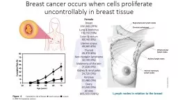
Breast cancer most frequently spreads by the hematogeneous route.
Veins of the breast
1. Internal mammary vein
2. perforating branches of IMV
3. intercostal vein
4. perforating branch of intercostal vein
5. lateral thoracic vein
6. axillary vein
7. subclavian vein.
Slide37The venous return from the breast
Slide38The Lymphatic System
The Lymphatic System transports a watery clear fluid called lymph. The lymphatic system aids the immune system in destroying pathogens and filtering waste so that the lymph can be safely returned to the circulatory system. The lymphatic system contains immune cells called lymphocytes, which protect the body against antigens such as harmful bacteria and viruses that invade the body.
Slide39Lymphatic System
Lymph flow begins deep within the breast tissues through lymphatic vessels that originate in the stroma and lactiferous ducts (deep system). Intramammary Lymph Nodes are seen throughout the breast parenchyma as part of the deep system.
Flow direction from the deep system is toward the areola into the periareolar plexus and continues into the subdermal lymphatic vessels (superficial system). From the subdermal vessels, lymph flows outward to the outer lymphatic chains that drain the breast. The outer lymphatic chains are located in multiple areas surrounding the breast.
Slide40Intra-mammary Lymph Node
Slide41Approximately 75% of lymphatic drainage is to the axilla. Therefore, the axillary lymph node chain becomes extremely important in predicting the spread of breast cancer.
The axillary lymph node chain consists of 6 groups of nodes:
1. External mammary group-located along the lateral thoracic vessels.
2. Scapular group-run with the subscapular vessels.
3. Axillary group- run with the axillary vessels.
4. Central group – run with the axillary vessels
5. Subclavicular group – run with the subclavian vessels.
6.Interpectoral (Rotter’s) nodes – found between pectoralis major and minor muscles.
Slide42Lymph nodes of the axillary region
Slide43The remaining 25% of lymphatic drainage include:
1. Internal mammary lymph nodes – lie along the internal mammary vessels.
2. Intercostal Lymph nodes
3. Flow to the opposite breast
4. Subclavicular Lymph nodes – Within the subclavicular fossa
5. Diaphragmatic Lymph Nodes – allow drainage to the abdomen.
Slide44The lymph node is reniform (kidney – like) in shape. It has an outer cortex and medial hilum where the small artery, vein, and lymph vessel enter and exit the node. Outer afferent lymphatic vessels carry lymph into the node where it is filtered and released through the hilar vessel.
Slide45Slide46Nerves
A complicated network of nerves serve the breast, chest muscles, an surrounding tissues.
These include:
1. Long thoracic nerve
2. thoraco-doral verve
3. Thoracic intercostal nerves
4. 3
rd
and 4
th
Branch of the cervical plexus
5
. Circumflex nerve (Axillary Nerve)
6. Subcapular nerves
7. Anterior thoracic nerves
Slide47Circumflex (Axillary) nerve
Slide48The nerves of the axilla region
Slide49Physiology and Hormonal Influences
Puberty
At puberty, breast development occurs due to hormonal stimulation by the ovaries (the larche).
The amount of growth of breast size and volume will vary with each individual.
Estrogen stimulates changes of stromal tissues: elongation of the mammary ducts, growth of connective tissue, increase in adipose tissue and increased vascularity.
Progesterone stimulates growth of the glandular tissue: TDLUs.
Slide50Mature female breast
The mature female breast is sensitive to the menstrual cycle and responds to fluctuating hormone levels every month. Early in the proliferative phase of the menstrual cycle, changes in the epithelium occur. Later in the secretory phase, the ducts and veins increase in size, the stroma becomes edematous, and the epithelium produces secretions. These changes may account for premenstrual breast discomfort. At the onset of menses, the breast tissues decrease in size.
Slide51Pregnancy
During pregnancy, there is considerable change in the breast tissue. The TDLUs increase in size as the epithelium begins to swell. The acinar cells enlarge in response to a variety of hormones including estrogen and progesterone, and lactogen, prolactin, and chorionic gonadrotrophin from the placenta.
Late in the pregnancy, the lactiferous ducts increase in size
.
Slide52Lactation
Shortly after birth, the estrogen and progesterone levels diminish rapidly and
Prolactin dominates
. This hormone causes the acinar cells to secrete milk.
After the termination of breast-feeding, the ducts and lobules return to their normal size in approximately 3 months.
Slide53Menopause
In the prei-menopausal female, the lobules of the breast involute (roll inward, invert). Also, the connective and stromal tissues are largely replaced by fat. Involuation is thought to begin long before menopause as a gradual decrease in glandular tissue with fatty replacement.
There are only a few situations, however, when breast parenchyma or glandular tissue increase with age.
The most common causes of increased glandular tissue development are:
pregnancy/lactation
Hormone replacement therapy (HRT)
Significant weight loss
The ratio of glandular to fatty tissue in the breasts is determined by
Total body fat : Total body weight
Slide54End modules one, two, three
Next week
Test on modules one, two, and three.