
1 Cheek Cells These cells were gently scraped from the inner surface of a persons cheek and placed on a microscope slide The cheek lining cells are thin and flat They fit together like tiles on a floor except that they overlap slightly Because they are thin and flat and several layers ID: 722509
Download Presentation The PPT/PDF document "Why are cells shaped the way they are?" is the property of its rightful owner. Permission is granted to download and print the materials on this web site for personal, non-commercial use only, and to display it on your personal computer provided you do not modify the materials and that you retain all copyright notices contained in the materials. By downloading content from our website, you accept the terms of this agreement.


Slide1
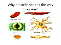
Why are cells shaped the way they are?Slide2
# 1 Cheek Cells
These cells were gently scraped from the inner surface of a person’s cheek, and placed on a microscope slide. The cheek lining cells are thin and flat. They fit together like tiles on a floor, except that they overlap slightly. Because they are thin and flat and several layers thick these cells make the lining of the cheek smooth, flexible, and strong. The large dark spot in the center of each cell is the nucleus. Slide3
# 1 Cheek Cells
Draw what you see
Label: the
nucleus
1) Why
are the cheek cells thin and flat? 2)Why do you think they need to fit together like tiles?Slide4
# 2 Blood Cells
The most numerous cells are red blood cells that look like doughnuts. They are flat disks, thinner in the middle than the edge, so it is light in the middle and darker on the edge. A red blood cell has no nucleus. It carries oxygen and carbon dioxide to and from body cells.
The white blood cells
(A and B)
are larger and have a nuclei. White blood cells help fight disease by removing foreign particles.Slide5
#2 Blood Cells
Draw what you see
Label: White blood cells and Red blood cells
1)What
is the function of red blood cells
?
2) Why are the red blood cells shaped like donuts? (Think of where they travel and what they carry in the center!)
A
BSlide6
# 4 Bone Cells
This is a very thin slice of bone, a living tissue. The two dark spots marked
“A”
are bone cells. There is a chemical that hardens in the space between cells.
Can you see many fine dark lines connecting each of the bone cells? Scientists believe these are canals that help chemicals go from cell to cell in the bones.
The dark area
(B) is a cross section of a tube. Blood vessels and nerves running through this tube bring food and oxygen to the bone cells.Slide7
# 4 Bone Cells
Draw what you see
Label: Bone Cell and Nerves/Blood Vessels
1) Why
do the bone cells look so rigid and strong?
2)Why
are blood cells so close to the bone cells? What do they help with?
A
A
BSlide8
# 5 Voluntary Muscle Cells
The long, spotted, ribbon-like shapes in this slide are voluntary muscle cell like in your arms and legs. These cells are very long.
Muscle cells contain many long fibers which can shorten and pull with great force. Many movements are caused by the contracting. Voluntary means under your control. At
“A”
you can see the nucleus. Because of their striped appearance, they are called striated. About 40% of the average person’s weight consist of voluntary muscle.Slide9
# 5 Voluntary Muscle Cells
Draw what you seeLabel: Nucleus
1) Why
are voluntary muscle cells so long? What do they do?
ASlide10
# 6 Involuntary Muscle Cells
This slide is made from a piece of the stomach wall. At
“A”
you can see a single voluntary muscle cell clearly with nucleus. Notice how much shorter the involuntary muscle is than the voluntary muscle. Involuntary muscle is located in the walls of the esophagus, stomach, and intestines. They are involuntary because they contract automatically without you thinking about it. They are not striated so scientists call them “smooth” muscle.Slide11
# 6 Involuntary Muscle Cells
Draw what you see
Label: Nucleus
1
)
Why are these cells so much shorter than voluntary muscle cells?2)Name two organs made of this type of muscle.
3)
What is the major difference between voluntary and involuntary muscles?
ASlide12
# 7 Nerve Cells
This tissue is taken from the spinal cord.
“A”
is the main part of the cell called the cell body. The cell has many long extensions which go very far in the body.
The ends of each branch make contact with other cells. Nerve cells are shaped like long wires so they can carry messages form one end of the body to the other.
Some nerve cells in the brain can keep their information and send out messages for a long time. This is how we learn and have memory.
“B” is a small blood vessel that brings food and oxygen to the nerve cells. Slide13
# 7 Nerve Cells
Draw what you see
Label: Cell Body, Nucleus, Branches, and Blood Vessel
1) Why
are nerve cells shaped the way they are?
2) Why
are we able to remember so much information in our brain?
3) What
does the blood vessel do for our nerve cells?
A
BSlide14
# 8 Gland Cells
This slide was made from a section of the wall of the large intestine. At
“A”
you can see a gland with its duct opening into the middle of the intestines. Around the duct are many glands cells.
“B”
is the nucleus of one of these cells. The gland cells produce a substance which burst form cells and enters the duct. The substance goes into the intestine and helps to break down food.Slide15
# 8 Gland Cells – (Gland cells secrete liquids needed in our body like digestive enzymes and hormones.)
Draw what you see
Label: Duct and Nucleus
1) What
is the function of gland cells?
2)Why do they have those pockets? What do they do?
A
B