
elorheostosisLeridiseaseisararenonhereditaryscle Fig1A46yearoldmanwithneckpainafterminortraumaAPandlateralradiographsshowadenselyscleroticrightparavertebralmassextendingfromtheC5toT1levelC ID: 944140
Download Pdf The PPT/PDF document "CASEREPORTMelorheostosisInvolvingtheCerv..." is the property of its rightful owner. Permission is granted to download and print the materials on this web site for personal, non-commercial use only, and to display it on your personal computer provided you do not modify the materials and that you retain all copyright notices contained in the materials. By downloading content from our website, you accept the terms of this agreement.

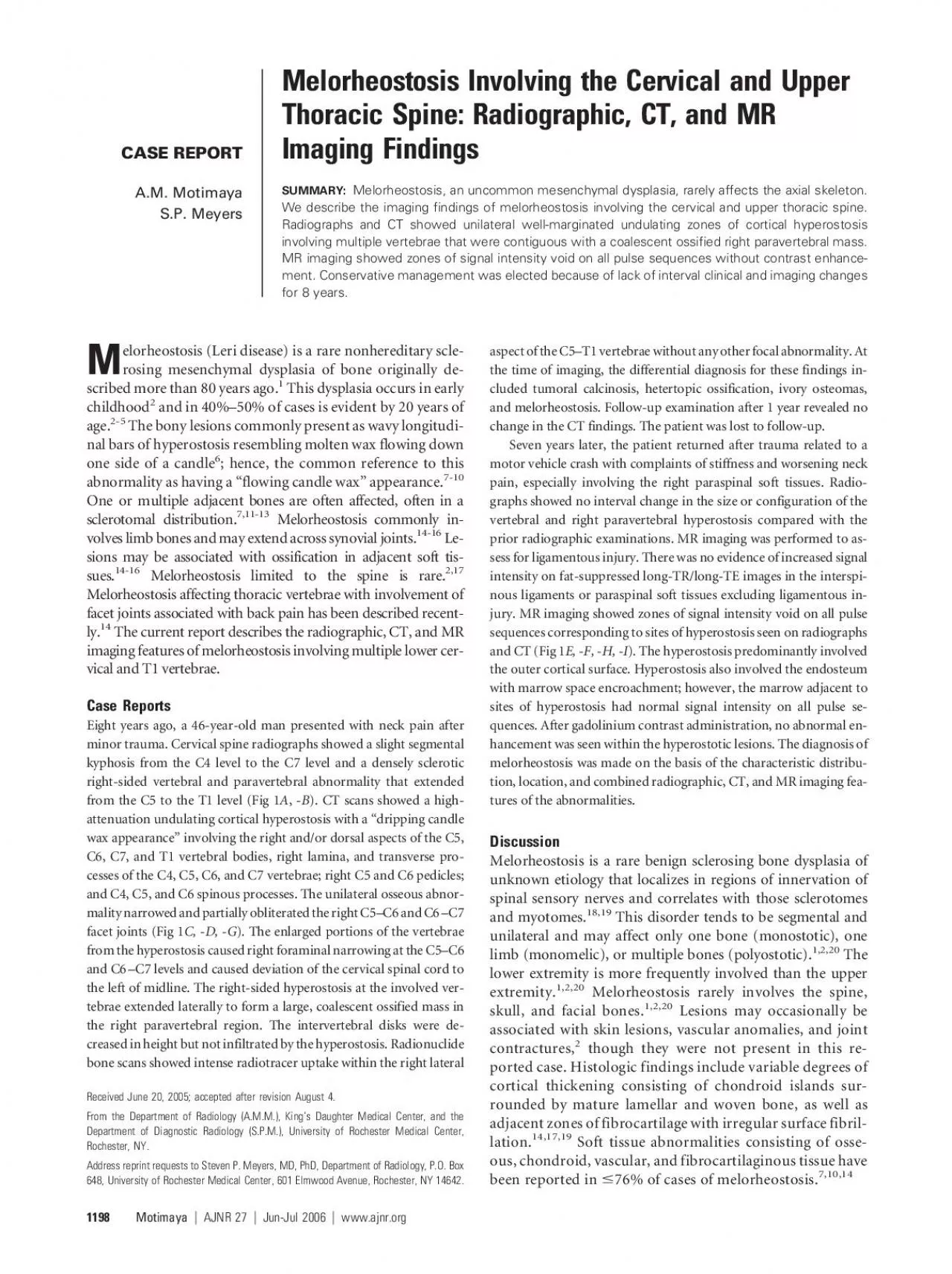
CASEREPORTMelorheostosisInvolvingtheCervicalandUpperThoracicSpine:Radiographic,CT,andMRImagingFindingsA.M.MotimayaS.P.MeyersMelorheostosis,anuncommonmesenchymaldysplasia,rarelyaffectstheaxialskeleton.Wedescribetheimagingfindingsofmelorheostosisinvolvingthecervicalandupperthoracicspine.RadiographsandCTshowedunilateralwell-marginatedundulatingzonesofcorticalhyperostosisinvolvingmultiplevertebraethatwerecontiguouswithacoalescentossifiedrightparavertebralmass.MRimagingshowedzonesofsignalintensityvoidonallpulsesequenceswithoutcontrastenhance-ment.Conservativemanagementwaselectedbecauseoflackofintervalclinicalandimagingchangesfor8years. elorheostosis(Leridisease)isararenonhereditaryscle- Fig1.A46-year-oldmanwithneckpainafterminortrauma.AP()andlateral()radiographsshowadenselyscleroticrightparavertebralmassextendingfromtheC5toT1level.Coronal(),rightparasagittal(),andaxial()CTimagesshowundulatingzonesofcorticalhyperostosiswithadrippingcandlewaxappearanceinvolvingtherightsidesoftheC5T1vertebraewithextensionovertherightfacetjointsofC4C5toC7T1.Thehyperostosisinvolvesthevertebralbodies,rightlaminaeandtransverseprocesses,andrightsidesofthespinousprocesses.Theexpansilecorticalhyperostosisextendsmostlyperipherallyalongtheouterbonesurfacebutalsoinvolvestheendostealmarginsresultinginprominentnarrowingoftheinvolvedmarrowspace.PosteriorvertebralbodyosteophytesatC3C4andnarrowingoftherightC5C6foramenfromthehyperostosisarealsoevident.Rightparasagittal()T1-weighted(TR450,TE9),rightparasagittal()T2-weighted(TR1800,TE110),axial)T2-weighted(TR4000,TE100),andaxial()postgadolinium-contrast(Gadoteridol)T1-weighted(TR450,TE9)MRimagesobtained7yearsaftertheradiographsandCTimagesaboveshowzonesofsignalintensityvoidonallpulsesequenceswithnoenhancementcorrespondingtotheareasofhyperostosisseenonradiographsandCTscans.AposteriorspondyloticridgeatC3C4()isseen.Sagittal,midline()T2-weighted(TR,1800;TE,110)MRimageshowstheposteriorspondyloticridgeatC3C4toresultinspinalstenosis,whichislikelysecondarytothefusedspinalcolumnfrommelorheostosisimmediatelybelowthislevel.Nosignalintensityabnormalities,however,areseeninthespinalcordrelatedtothespinalstenosisatC3C4orfromthemelorheostosisinvolvingthelowercervicallevels.CASEREPORTAJNRAmJNeuroradiol27:11981200Jun-Jul2006 Thedifferentialdiagnosisforsuperficialhyperattenuatedvertebralandadjacentparaspinalabnormalitiesincludesmelorheostosis,tumoralcalcinosis,tumoralcalciumpyro-phosphatedihydratedepositiondisease(CPPD),ivoryosteo-mas,heterotopicossification(myositisossificans),andparos-tealandperiostealosteosarcoma.Theunilateralandmultifocalcorticallocations,distinctimagingfeatures,andlackofintervalchangefortheabnormalitiesinthisreportare,however,characteristicformelorheostosisandareidenticaltopriorreportedimagingfindingsforbiopsy-confirmedmelo-rheostosisinvolvinglowerthoracicandlumbarvertebrae.Theimagingfeaturesofthiscasearealsosufficientlydifferentfromtheotherdisordersinthedifferentialdiagnosis.Forex-ample,tumoralcalcinosisofthespine,whichisoftenassoci-atedwit
hsystemicdisordersofcalciummetabolismorrenaldialysis,typicallyoccursashighattenuationparaosseousle-sionsthatresultfromdystrophiccalcificationsinsofttissuescomposedofcalciumhydroxyapatitecrystals,collagenousfi-broussepta,andcollectionsofhistiocytesandforeignbodygiantcells.Unlikemelorheostosis,tumoralcalcinosisofthespineisoftennotunilateralandappearsprimarilyasex-tradurallesionswithheterogeneousmixedsignalintensityonbothT1-weightedandT2-weightedimages,whichareoftenassociatedwitherosionofadjacentbone.CPPDcanoccurastumor-likelesionsinthespine,typicallylocatedattheliga-mentumflavumandsynovialjoints.CPPDlesionscontainneedle-andrhomboid-shapedcalciumpyrophosphatecrys-talswithassociatedchondroidelements,fibrocollageneoustissue,andvariableamountsofacuteandchronicinflamma-toryreaction.CPPDlesionsmaycauseerosionofadjacentTheMRimagingfindingsofspinalCPPDarealsosimilartothosefortumoralcalcinosis,thusdifferingfromthoseformelorheostosis.Althoughivoryosteomascanhavehistologicfeaturessimilartomelorheostosis,largeosteomasofthespineareveryrareandhavebeenreportedtoinvolveonlysinglevertebra.Heterotopicossification,alsoreferredtoasmyositisossificans,typicallyoccursaslesionsinsofttissuewithpredominantperipheraldistributionsofossi-ficationandthusdiffersfromtheappearanceofcortical-basedhyperostosisseenwithmelorheostosis.Theimagingfeaturesofparostealandperiostealosteosarcomasalsodiffermarkedlyfrommelorheostosisbecausethesetumorscontainossificmineralizedmatrixthatisoftenirregularandnotuniformlyattenuatedonradiographsandhavefocalorpoorlydefinedsoft-tissuemasseswithhighsignalintensityonT2-weightedTumorinvasionintomedullarybonewithhighsignalintensityonT2-weightedimagesoccurin41%oflow-gradeand50%ofhigh-gradeparostealosteosarcomas.nally,thejuxtacorticalsofttissuemassesofperiostealosteo-sarcomastypicallycauseextrinsicerosionofcorticalbone,perpendicularperiostealreaction,andreactivemarrowzoneswithhighsignalintensityonT2-weightedimages,whicharefindingsnotseenwithmelorheostosis.Althoughradiographicandbonescintigraphicappearanceofmelorheostosishasbeenwelldescribed(ie,undulatingcorticalthickeningandmarkedincreaseduptakeofradionuclide),CTandMRimaginghelpsconfirmandaccuratelylocalizethezonesofhyperostosisinthespineandprovideassessmentofthedegreesofnarrowingofthespinalcanalandforamina.Further,eventhoughtheMRimagingappearanceofsofttissuemassesassoci-atedwithmelorheostosisisvariable,mineralizedandnonminer-alizedsofttissueabnormalitiesshouldberecognizedasanothermanifestationofthisdisease.MRimagingaidsinconfirmationofthediagnosisandintheaccuratedetectionanddetermi-nationoftheextentofsofttissueinvolvement.Althoughmelorheostosisisarareconditionaffectingtheaxialskeleton,itshouldbeadefiniteconsiderationinthedif-ferentialdiagnosisofunilateralorsegmentallesionsofcorticalhyperostosisinthespinebecauseaccuratedetectioncanpre-ventanunwarrantedbiopsy.1.LeriA,JoannyJ.Uneaffectionnonde´critedesoshyperostoseencoule´esurtoutelalongeurdunmemberoumelorhe´ostose.BullMemoiresSocMedHopitauxParis1922;46:1141452.Kalbermat
tenNT,VockP,Ru¨fenachtD,etal.Progressivemelorheostosisintheperipheralandaxialskeletonwithassociatedvascularmalformations:im-agingfindingsoverthreedecades.SkeletalRadiol2001;30:48523.IsaacsP,ResnickD.Lettertotheeditors:MRappearanceofaxialmelorheos-SkeletalRadiol1993;22:47484.YoungeD,DrummondD,HerringJ,etal.Melorheostosisinchildren:clinicalfeaturesandnaturalhistory.JBoneJointSurgAm5.BrownRR,SteinerGC,LehmanWB.Melorheostosis:casereportwithradio-logic-pathologiccorrelation.SkeletalRadiol2000;29:548526.SoffaDJ,SireDJ,DodsonJH.Melorheostosiswithlinearsclerodermatousskin7.JudkiewiczAM,MurpheyMD,ResnikCS,etal.Advancedimagingofmelo-rheostosiswithemphasisonMRI.SkeletalRadiol8.CampbellCJ,PapademetriouT,BonfiglioM.Melorheostosis:areportoftheclinical,roentgenographic,andpathologicalfindingsinfourteencases.JBoneJointSurgAm9.MorrisJM,SamilsonRL,CorleyCL.Melorheostosis:reviewoftheliteratureandreportofaninterestingcasewithanineteenyearfollow-up.JBoneJointSurgAm10.MurrayRO,McCredieJ.Melorheostosisandthesclerotomes:aradiologicalSkeletalRadiol11.BaerSC,AyalaAG,RoJY,etal.Casereport843.SkeletalRadiol1994;23:3101412.SiegelA,WilliamsH.Casereports:linearsclerodermaandmelorheostosis.BrJRadiol1992;65:2666813.GoldmanAM,SchneiderR,HuvosAS,etal.Casereport778.SkeletalRadiol1993;22:20621014.McCarthyM,MehdianH,FairbairnKJ,etal.Melorheostosisofthetenthandelevenththoracicvertebraecrossingthefacetjoint:ararecauseofbackpain.SkeletalRadiol2004;33:2838615.GarverP,ResnickD,HaghighiP,etal.Melorheostosisoftheaxialskeletonwithassociatedfibrolipomatouslesions.SkeletalRadiol1982;9:414416.GreenspanA,AzouzEM.Bonedysplasiaseries:melorheostosis:reviewandCanAssocRadiolJ1999;50:3243017.RobertsonPA,DonAS,MillerMV.Painfullumbosacralmelorheostosistreatedbyfusion.2003;28:E2343818.ResnickD,NiwayamaG.Enostosis,hyperostosis,andperiostitis.In:ResnickE,ed.Diagnosisofboneandjointdisorders.3rded.Vol6.Philadelphia:Saunders;1995:44101519.ZeillerSC,VaccaroAR,WimberleyDW,etal.Severemyelopathyresultingfrommelorheostosisofthecervicothoracicspine.Acasereport.JBoneJointSurgAm2005;87:27956220.KhuranaJS,EharaS,RosenbergAE,etal.Melorheostosisofilium,femur,andadjacentsofttissues.SkeletalRadiol21.DurantDM,RileyLH,BurgerPC,etal.Tumoralcalcinosisofthespine,astudyof21cases.22.KokubunS,OzawaH,SakuraiM,etal.Tumoralcalcinosisintheuppercervicalspine:acasereport.1996;21:2495223.OhashiK,YamadaT,IshikawaT,etal.Idiopathictumoralcalcinosisinvolvingthecervicalspine.SkeletalRadiol1996;25:3889024.ShaffreyCI,MunozEL,SuttonCL,etal.Tumoralcalciumpyrophosphatedehydratedepositiondiseasemimickingacervicalspineneoplasm:casere-25.RengacharySS,SananA.Ivoryosteomaofthecervicalspine:casereport.1998;42:1828526.EharaS,ShiraishiH,AbeM,etal.Reactiveheterotopicossification,itspatternsonMRI.ClinImaging27.JelinekJS,MurpheyMD,KransdorfMJ,etal.Parostealosteosarcoma:valueofMRimagingandCTinthepredictionofhistologicgrade.8374228.MurpheyMD,JelinekJS,TempleHT,etal.Imagingofperiostealosteosarcoma:radiologic-pathologiccomparison.2004;233:12938AJNR27Jun-Jul2006