
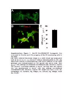
Figure S3 MILI antibody specifically recognizes MILImilimilimili testes Tubulin serveseIF3a eIF4GII and ribosomal protein S6 in MILI antibody coprecipitated MILI and eIF3a from ID: 824702
Download Pdf The PPT/PDF document "SUPPLEMENTARY FIGURE LEGNEDS Figure S1. ..." is the property of its rightful owner. Permission is granted to download and print the materials on this web site for personal, non-commercial use only, and to display it on your personal computer provided you do not modify the materials and that you retain all copyright notices contained in the materials. By downloading content from our website, you accept the terms of this agreement.

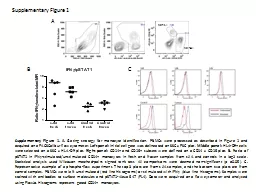


SUPPLEMENTARY FIGURE LEGNEDS Figure S1.
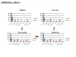
SUPPLEMENTARY FIGURE LEGNEDS Figure S1. The defect of mutant in germline stem cell self-renewal.stained with hematoxylin and eosin and shown under the same magnification for easy comparison. sg: spermatogonia; sc: spermatocytes; st: spermatids; stc:
Sertoli cells. Further characterizatio
Sertoli cells. Further characterization of germlinetestes (A) and (B) mice stained for TUNNEL (red), laminin (green), spermatogonia +/-mice stained for MILI (red), Sertoli cells (by anti-TSX antibody, green), and DNA (blue). Sertoli cells are pr
esent in the mutant testis but MIe this
esent in the mutant testis but MIe this is a protein-null allele. (E) and (F) mice stained for BC7 antibody and DAPI, which label primary spermatocytes (green) and DNA (blue). A small number of germ cells in the mutant testis have differentiated
into spermatocytes, as also testicular
into spermatocytes, as also testicular sections of six-month old -/- (H) mice stained for EE2 to label spermatogonia (red), phosphohistone 3 to label mitotic chromosomes (green), and DNA (blue). In the testis, many spermatogonia and spermatocytes
are undergoing active mitosis, yet in th
are undergoing active mitosis, yet in the mitosis is not detectable. testicular sections of six-month old (H) mice stained for laminin (green) to outline seminiferous tubules, TUNNEL labeling to identify detectable, yet the Figure S3. MILI antibo
dy specifically recognizes MILI.mili(+/+
dy specifically recognizes MILI.mili(+/+)mili(+/-mili(-/-) testes. Tubulin serveseIF3a, eIF4GII, and ribosomal protein S6 in MILI antibody co-precipitated MILI and eIF3a from the 8dpp testicular extract. The co-immunoprecipitatiILIeIF4GIIS68dppAF