
Blouin Y Cazajous G Dehan C Soler C Vong R Hassan M et al Progenitor Mycobacterium canettii Clone Responsible for Lymph Node Tuberculosis Epidemic Djibouti Emerg Infect Dis 20142012128 httpsdoiorg103201eid2001130652 ID: 1043883
Download Presentation The PPT/PDF document "Figure 2 Figure 2. . Early evolution of ..." is the property of its rightful owner. Permission is granted to download and print the materials on this web site for personal, non-commercial use only, and to display it on your personal computer provided you do not modify the materials and that you retain all copyright notices contained in the materials. By downloading content from our website, you accept the terms of this agreement.


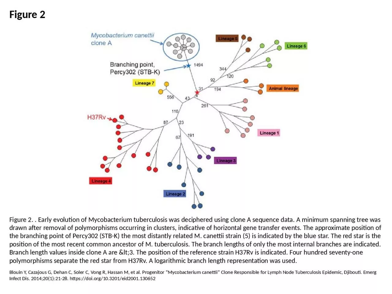
1. Figure 2Figure 2. . Early evolution of Mycobacterium tuberculosis was deciphered using clone A sequence data. A minimum spanning tree was drawn after removal of polymorphisms occurring in clusters, indicative of horizontal gene transfer events. The approximate position of the branching point of Percy302 (STB-K) the most distantly related M. canettii strain (5) is indicated by the blue star. The red star is the position of the most recent common ancestor of M. tuberculosis. The branch lengths of only the most internal branches are indicated. Branch length values inside clone A are <3. The position of the reference strain H37Rv is indicated. Four hundred seventy-one polymorphisms separate the red star from H37Rv. A logarithmic branch length representation was used.Blouin Y, Cazajous G, Dehan C, Soler C, Vong R, Hassan M, et al. Progenitor “Mycobacterium canettii” Clone Responsible for Lymph Node Tuberculosis Epidemic, Djibouti. Emerg Infect Dis. 2014;20(1):21-28. https://doi.org/10.3201/eid2001.130652