
Presented by DrKANBARASAN Assistant professor and Head Department of Botany Padmavani Arts and Science College for Women Salem 636 011 ELECTROPHORESIS Electrophoresis is a molecular technique widely used in field of biochemistry molecular biology and biotechnology to separate the ID: 1032810
Download Presentation The PPT/PDF document "Protein separation by SDS – PAGE" is the property of its rightful owner. Permission is granted to download and print the materials on this web site for personal, non-commercial use only, and to display it on your personal computer provided you do not modify the materials and that you retain all copyright notices contained in the materials. By downloading content from our website, you accept the terms of this agreement.


1. Protein separation by SDS – PAGE Presented byDr.K.ANBARASANAssistant professor and HeadDepartment of Botany Padmavani Arts and Science College for WomenSalem - 636 011.
2. ELECTROPHORESISElectrophoresis is a molecular technique widely used in field of biochemistry, molecular biology and biotechnology to separate the bio molecules based on the migration of charged molecules.The rate of migration depends on the strength of electric field, size and shape of the molecules.
3. PrincipleWhen charged molecules are placed in an electric field they migrate towards either the positive (anode) or negative (cathode) pole according to their charge.
4. Factors affected electrophoresis mobilityNet charge of the moleculeSize and shapeConcentration of molecule in solutionElectric strength
5. Electrophoresis unitElectrodesBuffer tanks or chambersSupporting mediumSpacer & combTransparent insulating glass platesDC power supply
6. Electrophoresis MediaBufferSupporting mediumBuffer: Solution of week acids or saltsFunctions of buffer1. carries the applied current2. determine the electric charge on the solute3. Maintains the pHCommonly used bufferPhosphate bufferTris borate bufferTris glycine buffer
7. Supporting mediumFeatures of supporting mediaChemical nature – Inert Availability – Easy Transparency _ HighAdsorptivity – Low Sieving effect – desirablePreparation – EasyPorosity – ControlledElectrical conductivity – HighCommonly used supporting mediaPaper(Cellulose) – Poor conductor of electricity e.g. What mann paper Cellulose acetate – It used to separation of clinical medicineStarch – It used for separation of some iso- enzymesAgarose – Agarose is purified form of agar, it is polysaccharide used for nucleic acid separation.Polyacrylamide gel – It is inert and highly transparent and also used for protein and amino acid separation.
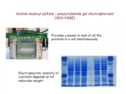
8. SDS-PAGE (Sodium Dodecyl Sulfate – Polyacrylamide gel Electrophoresis )Materials needed:Vertical electrophoresis apparatusPower pack30% Acrylamide mix (29% Acrylamide and 1% Bis-acrylamide)1.5M Tris (pH 8.8)1M Tris (pH6.8)10% SDS10%APS (Freshly prepared)TEMED(N,N,N’,N’-Tetra methylene diamine)Tris-Glycine electrophoresis bufferStaining solutionDistaining solutionDistilled water
9. Role of ReagentsPolyacrylamide, as medium for vertical gel electrophoresis it composed of a mixture of two chemicals: acrylamide and bis-acrylamide.Acrylamide forms long polymer chains. Bisacrylamide is cross linking agent and links long polymers of acrylamide. Both these components will form a cross linked network in the presence of APS called polymerization.APS (Ammonium Persulfate) is polymerizing agent which generate free radicals(charged oxygen) in the presence of TEMED (is a free radical stabilizer)Air inhibits polymerization as it scavenges free radicalsPore size is determined by % of acrylamide and the amount of cross linker.
10. SDS SDS is used in the gel mix.SDS is –ve charged and binds to proteins, it denatures (unfolds) proteins and gives a net negative charge. Proteins will then migrate to the anode.Proteins all have same charge to mass ratioCan be separated based on size aloneBiomolecules moved with a speed dependent on their charge, shape, and size and separation occures on the basis of molecular size.
11. Stacking gelThis gel has a large pore size and a lower pH than the separating gel so the protein sample are easily pass through the gel.Separating gel or resolving gelAs the proteins migrate through the resolving gel, the larger proteins will move more slowly and the proteins will be separated by size. Stacking gel (4%)Separating gel (12.5%)Acrylamide -bisacrylamid(30%)850µlAcrylamide -bisacrylamid(30%)2.08 ml0.5M Stacking buffer(pH 6.8)625 µl1.5M Separating buffer(pH 8.8)1.25 ml10% SDS50 µl10% SDS50 µl10% APS50 µl10% APS50 µlTEMED5 µlTEMED2 µlDistilled water3.45mlDistilled water1.61 ml
12. Staining solutionCoomassie brilliant blue (Dissolve 15mg coomassie brilliant blue in 0.2ml methanol and add 9.8ml of distilled waterDestaining solution - IMethanol - 375mlAcetic acid - 35mlDistilled water - 215mlDestaining solution – IIMethanol - 250mlAcetic acid - 35mlDistilled water - 215ml
13. ProcedureAssemble the glass platesPrepare acrylamide solutions (Stacking and separating gels)Pour the separating gel into the glass plate and keep in room temperatureKeep for polymerization about 30 – 40 minutes (After 30minute is complete polymerization)Pour the stacking gel solution directly on to the surface of the polymerized resolving gelImmediately insert a clean comb in to the gel solution
14. While the stacking gel is polymerizing and prepare required volume of protein samplesHeat the samples in boiling water both for 2min to denature the proteins. Keep them on ice to retain the denature state.Add electrophoresis buffer to the top and bottom buffer chambers of the electrophoresis apparatus.Remove the comb carefully, after the polymerization and load the sample along with marker proteins into the wells.
15. Attach the apparatus to power supply unit and apply electric power of 50 to 100VRun the gel until bromophenol blue dye reaches the bottom of resolving gel
16. Turn of power supply and remove the glass plate from the electrophoresis apparatusThe gel was immersed at least 100ml of staining solution (Coomassie blue) and kept overnight at room temperature.The gel was then destained by soaking it in the destaining solution for every 2 hours, the destaining solution was changed and it was repeated to about 3 – 4 timesAfter destaining; the gel was stored in 20% glycerol in a sealed plastic bagVisualize the bonds in an illuminator and take the photographs.The molecular weight of the bands seen on the gel was indicated by the position of the molecular weight marker protein.
17. Thanking you.,