
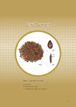
217Figure 1 A Lini Semen217121 cm1 mm1 mmACBLini SemenLini Semen2181Chinese Phonetic Name Yamazi2SOURCELini Semen is the dried ripe seed of Linum usitatissimum L Linaceae The plant is collected in a ID: 870715
Download Pdf The PPT/PDF document "A photograph of Lini Semen" is the property of its rightful owner. Permission is granted to download and print the materials on this web site for personal, non-commercial use only, and to display it on your personal computer provided you do not modify the materials and that you retain all copyright notices contained in the materials. By downloading content from our website, you accept the terms of this agreement.


1 217 Figure 1 A photograph of Lini Sem
217 Figure 1 A photograph of Lini Semen A. Lini Semen 217 1 2 1 cm 1 mm 1 mm A C B Lini Semen Lini Semen 218 1. Chinese Phonetic Name: Yamazi 2. SOURCE Lini Semen is the dried ripe seed of Linum usitatissimum L. (Linaceae). The plant is collected in autumn when the seed is ripe, the harvested plant dried under the sun, the seeds tapped out, foreign 3. Ovoid, �at, 2.3-5.9 mm long, 1.6-2.9 mm wide, 0.7-1.3 mm thick. Externally brown to reddish-brown, smooth and lustrous. One end acuminate and slightly oblique, the other end obtuse, with raphe and dented hilum on the acuminate end. Testa and endosperm thin, cotyledons 2, yellowish-white, oily. IDENTIFICATION 4.1 Microscopic (Appendix III) Transverse Epidermis of testa consists of 1 layer of rectangular cells with mucilaginous walls. Hypodermis consists of 1 layer of cells, walls slightly thickened. Sclerenchymatous cells layer consists of 1 layer of densely arranged cells, walls thickened. Parenchymatou
2 s cells layer consists of collapsed cel
s cells layer consists of collapsed cells, boundaries indistinct. Pigment layer consists of 1 layer of �attened cells �lled with pigment masses. Endosperm consists of several layers of polygonal cells filled with aleurone grains. Powder Colour yellowish-brown to pale brown. Pigment cells pale yellow to yellow, square, rectangular or polygonal in surface view, 9-49 µm in diameter, anticlinal walls �nely beaded, always containing yellowish-brown, brown or reddish-brown masses. Pigment masses yellowish-brown, brown or reddish-brown, square, rectangular or polygonal, edges usually finely serrated. Epidermal cells of testa large, polygonal in surface view, 16-69 µm in diameter, with mucilaginous walls. Sclerenchymatous cells pale yellow to yellow, strip-shaped, 4-23 µm in diameter, walls thickened or slightly thickened, with fine and dense pits; yellow or white under the polarized microscope. Hypodermal cells colourless to pale yello
3 w, subpolygonal to subrounded in surface
w, subpolygonal to subrounded in surface view, walls slightly thickened. Endosperm cells polygonal to subpolygonal, wall slightly thickened, �lled with aleurone grains and oil droplets. Cotyledon cells polygonal to subpolygonal, relatively small, �lled Figure 2 C B A 1 1 2 7 2 7 2 Lini Semen 221 Figure 3 1a 7a 2a Lini Semen 222 4.2 Thin-LayerChromatographic [Appendix IV(A)] Linoleic acid standard solution Weigh 0.2 mg of linoleic acid CRS (Fig. 4) and dissolve in 1 mL of ethanol. α-Linolenic acid standard solution Weigh 0.2 mg of α-linolenic acid CRS (Fig. 4) and dissolve in 1 mL of ethanol. reagent Add slowly 5 mL of sulphuric acid to 95 mL of ethanol. Test Weigh 0.2 g of the powdered sample and place it in a 100-mL conical �ask, then add 50 mL of n -hexane. Sonicate (300 W) the mixture in a water bath at about 60ºC for 1 h. Filter and transfer the filtrate to a 100-mL round-bottomed flask. Evaporate the solvent to dryness at reduced
4 pressure in a rotary evaporator. Disso
pressure in a rotary evaporator. Dissolve the residue in 5 mL of isopropanol. Filter through a 0.45-µm PTFE �lter. Procedure Carry out the method by using a HPTLC RP-18 F plate, a twin trough chamber and a freshly prepared developing solvent system as described above. Apply separately linoleic acid standard solution (3.5 μL), α-linolenic acid standard solution (2.5 μL) and the test solution (6 μL) to the r with a lid and let equilibrate for about 15 min. Carefully tilt the chamber to allow suf�cient solvent to pass from the trough containing the solvent to the other containing the HPTLC plate for development. Develop over a path of about 8 cm. After the development, remove the plate from the chamber, mark the solvent front and dry in air. Spray the plate evenly with the spray reagent and heat at about 105ºC (about 3 - 5 min). Examine the plate under UV light (366 nm). Calculate the R f values by using the equation as indicated in Ap
5 pendix IV (A). Figure 4 Chemical structu.
Figure 4
Chemical structu")
pendix IV (A). Figure 4 Chemical structures of (i) linoleic acid (ii) α-linolenic acid and (iii) secoisolariciresinol 1,4-diglucoside HOO ii iii HOO Figure 5 A reference HPTLC chromatogram of Lini Semen extract observed under UV light (366 nm) Test solution For positive identification, the sample must give spots or bands with chromatographic characteristics, including the colour and the R f values, corresponding to those of linoleic acid and 4.3 High-PerformanceChromatographic (Appendix XII) Linoleic acid standard solution for �ngerprinting, Std-FP (30 mg/L) Weigh 0.3 mg of linoleic acid CRS and dissolve in 10 mL of ethanol. α-Linolenic acid standard solution for �ngerprinting, Std-FP (40 mg/L) Weigh 0.4 mg of α-linolenic acid CRS and dissolve in 10 mL of ethanol. Test Weigh 0.2 g of the powdered sample and place it in a 50-mL centrifuge tube, then add 50 mL of n -hexane. Sonicate (300 W) the mixture in a water bath at about 60ºC for 1 h.
6 Centrifuge at Start about 4000 × f
Centrifuge at Start about 4000 × for 10 min. Filter and transfer the filtrate to a 250-mL round-bottomed flask. Repeat the extraction for two more times. Combine the �ltrates. Evaporate the solvent to dryness at reduced pressure in a rotary evaporator. Dissolve the residue in isopropanol. Transfer the solution to a 25-mL of volumetric �ask and make up to the mark with isopropanol. Filter through a 0.45-µm PTFE �lter. Chromatographic The liquid chromatograph is equipped with a DAD (210 nm) and a column (2.1 × 100 mm) packed with OS bonded silica gel (3.5 µm particle size). The column temperature is maintained at 40ºC during the separation. The �ow rate is about 0.6 mL/min. Programme the chromatographic system as follows (Table 1) – Table Chromatographic system conditions requirements Perform at least �ve replicate injections, each using 10 µL of linoleic acid Std-FP and α-linolenic acid Std-FP. The r
7 equirements of the system suitability pa
equirements of the system suitability parameters are as follows: the RSD of the peak areas of linoleic acid and α-linolenic acid should not be more than 5.0%; the RSD of the retention times of linoleic acid and α-linolenic acid peaks should not be more than 2.0%; the column ef�ciencies determined from linoleic acid and α-linolenic acid peaks should not be less R value between peak 1 and the closest peak; and the R value between peak 2 and the closest Procedure Separately inject linoleic acid Std-FP, α-linolenic acid Std-FP and the test solution (10 µL each) into the HPLC system and record the chromatograms. Measure the retention times of linoleic acid and α-linolenic acid peaks in the chromatograms of linoleic acid Std-FP, α-linolenic acid Std-FP and the retention times of the six characteristic peaks (Fig. 6) in the chromatogram of the Time Water Isopropanol test solution. Identify linoleic acid and α-linolenic acid peaks in the chromatogram
8 of the test solution by comparing its r
of the test solution by comparing its retention time with that in the chromatograms of linoleic acid Std-FP and α-linolenic acid Std-FP. The retention times of linoleic acid and α-linolenic acid peaks in the chromatograms of the test solution and the corresponding Std-FP should not differ by more than 2.0%. Calculate the RRTs of the characteristic peaks by using the equation as indicated in The RRTs and acceptable ranges of the six characteristic peaks of Lini Semen extract are listed in Table 2. Table The RRTs and acceptable ranges of the six characteristic peaks of Lini Semen extract Figure 6 A reference �ngerprint chromatogram of Lini Semen extract For positive identi�cation, the sample must give the above six characteristic peaks with RRTs falling within the acceptable range of the corresponding peaks in the reference fingerprint RRT 1 (marker, α-linolenic acid) 1.11 2.77 2.87 227 5. 5.1 Heavy (Appendix V) 5.2Pesticide Residues (Appen
9 dix VI) : meet the requirements. 5.3 Myc
: meet the requirements.
5.3 Myc")
dix VI) : meet the requirements. 5.3 Mycotoxins (Appendix VII) 5.4 Sulphur (Appendix XVI) : meet the requirements. 5.5 Foreign (Appendix VIII) : not more than 2.0%. 5.6 (Appendix IX) Total ash: not more than 4.0%. 5.7 Water (Appendix X) 6. (Appendix XI) Water-soluble extractives (cold extraction method): not less than 16.0%. ASSAY 7.1 Assay Carry out the method as directed in Appendix IV (B). Mixed linoleic acid and α-linolenic acid standard stock solution, Std-Stock (320 mg/L for linoleic acid and 400 mg/L for α-linolenic acid) Weigh accurately 1.6 mg of linoleic acid CRS and 2.0 mg of α-linolenic acid CRS, and dissolve in 5 mL of ethanol. 228 Mixed linoleic acid and α-linolenic acid standard solution for assay, Std-AS Measure accurately the volume of the mixed linoleic acid and α-linolenic acid Std-Stock, dilute with ethanol to produce a series of solutions of 5, 10, 20, 40, 80 mg/L for linoleic acid and 6.25, 12.5, 25, 50, 100 mg/L for α-linolenic
10 acid. Test Weigh accurately 0.2 g of th
acid. Test Weigh accurately 0.2 g of the powdered sample and place it in a 50-mL centrifuge tube, then add 50 mL of n -hexane. Sonicate (300 W) the mixture in a water bath at about 60ºC for 1 h. Centrifuge at about 4000 × for 10 min. Filter and transfer the filtrate to a 250-mL round- bottomed �ask. Repeat the extraction for two more times. (Collect the residue for the assay of secoisolariciresinol 1,4-diglucoside.) Combine the filtrates. Evaporate the solvent to dryness at reduced pressure in a rotary evaporator. Dissolve the residue in isopropanol. Transfer the solution to a 25-mL of volumetric �ask and make up to the mark with isopropanol. Filter through a 0.45-µm PTFE �lter. Chromatographic The liquid chromatograph is equipped with a DAD (210 nm) and a column (2.1 × 100 mm) packed with OS bonded silica gel (3.5 µm particle size). The column temperature is maintained at 40ºC during the separation. The �ow rate
11 is about 0.6 mL/min. Programme the chrom
is about 0.6 mL/min. Programme the chromatographic system as follows (Table 3) – Table Time Water Isopropanol requirements Perform at least five replicate injections, each using 10 µL of the mixed linoleic acid and α-linolenic acid Std-AS (20 mg/L for linoleic acid and 25 mg/L for α-linolenic acid). The requirements of the system suitability parameters are as follows: the RSD of the peak areas of linoleic acid and α-linolenic acid should not be more than 5.0%; the RSD of the retention times of linoleic acid and α-linolenic acid peaks should not be more than 2.0%; the column efficiencies determined from linoleic acid and α-linolenic acid peaks should not be less than R R value between α-linolenic acid peak and the closest peak in the chromatogram of the test solution should not be less than 1.5 Inject a series of the mixed linoleic acid and α-linolenic acid Std-AS (10 µL each) into the HPLC system and record the chromatograms. Plot the peak areas of lin
12 oleic acid and α-linolenic acid agains
oleic acid and α-linolenic acid against the corresponding concentrations of the mixed linoleic acid and α-linolenic acid Std-AS. r 2 Procedure Inject 10 µL of the test solution into the HPLC system and record the chromatogram. Identify linoleic acid and α-linolenic acid peaks (Fig. 7) in the chromatogram of the test solution by comparing their retention times with those in the chromatogram of the mixed linoleic acid and α-linolenic acid Std-AS. The retention times of linoleic acid and α-linolenic acid peaks in the chromatograms of the test solution and the Std-AS should not differ by more than 5.0%. Measure the peak areas and calculate the concentrations (in milligram per litre) of linoleic acid and α-linolenic acid in the test solution, and calculate the percentage contents of linoleic acid Appendix IV (B). The sample contains not less than 0.56% of the total content of linoleic acid (C 18 O 2 ) and 18 O 2 ), calculated with reference to the dried substance
13 . Figure 7 A reference assay chromatogr
. Figure 7 A reference assay chromatogram of linoleic acid and α-linolenic acid of Lini Semen extract 7.2 Assaysecoisolariciresinol Carry out the method as directed in Appendix IV (B). Secoisolariciresinol 1,4-diglucoside standard stock solution, Std-Stock (720 mg/L) Weigh accurately 3.6 mg of secoisolariciresinol 1,4-diglucoside CRS (Fig. 4) and dissolve in 5 mL of methanol (70%). Secoisolariciresinol 1,4-diglucoside standard solution for assay, Std-AS Measure accurately the volume of the secoisolariciresinol 1,4-diglucoside Std-Stock, dilute with methanol (70%) to produce a series of solutions of 4.5, 9, 18, 36, 72 mg/L for secoisolariciresinol Test Weigh accurately 0.2 g of the powdered sample and place it in a 50-mL centrifuge tube, then add 50 mL of n -hexane. Sonicate (300 W) the mixture in a water bath at about 60ºC for 1 h. Centrifuge at about 4000 × for 10 min. Filter and transfer the �ltrate to a 250-mL round-bottomed �ask. Repeat
14 the extraction for two more times. (Col
the extraction for two more times. (Collect the �ltrates for the assay of linoleic acid and α-linolenic acid.) Dry the residue for 2 h. Transfer the dried residue to a 50-mL conical �ask, then add 20 mL of sodium hydroxide (4%, w/v) in methanol (70%). Cap the �ask. Sonicate (300 W) the mixture in a water bath at about 60ºC for 1 h. Transfer the mixture to a 50-mL centrifuge tube and centrifuge at about 4000 × for 10 min. Filter and transfer the �ltrate to a 250-mL conical �ask. Repeat the extraction for one more time. Combine the �ltrates and adjust the pH to 3 with hydrochloric acid (4.37%, w/v) in methanol (70%). Transfer the solution to a 100-mL volumetric �ask and make up to er. Chromatographic The liquid chromatograph is equipped with a DAD (280 nm) and a column (4.6 × 250 mm) packed with ODS bonded silica gel (5 µm particle size). The column temperature is maintained at 30ºC
15 during the separation. The flow rate is
during the separation. The flow rate is about 1.0 mL/min. Programme the chromatographic system as follows (Table 4) – Table Time Water requirements Perform at least five replicate injections, each using 10 µL of secoisolariciresinol 1,4-diglucoside Std-AS (18 mg/L). The requirements of the system suitability parameters are as follows: the RSD of the peak area of secoisolariciresinol 1,4-diglucoside should not be more than 5.0%; the RSD of the retention time of secoisolariciresinol 1,4-diglucoside peak should not be more than 2.0%; the column ef�ciency determined from secoisolariciresinol 1,4-diglucoside peak should not be less than 25000 R value between secoisolariciresinol 1,4-diglucoside peak and the closest peak in the Inject a series of secoisolariciresinol 1,4-diglucoside Std-AS (10 µL each) into the HPLC system and record the chromatograms. Plot the peak areas of secoisolariciresinol 1,4-diglucoside against the corresponding concentrations of
16 secoisolariciresinol 1,4-diglucoside Std
secoisolariciresinol 1,4-diglucoside Std-AS. Obtain the slope, r 2 Procedure Inject 10 µL of the test solution into the HPLC system and record the chromatogram. Identify secoisolariciresinol 1,4-diglucoside peak (Fig. 8) in the chromatogram of the test solution by comparing its retention time with that in the chromatogram of secoisolariciresinol 1,4-diglucoside Std-AS. The retention times of secoisolariciresinol 1,4-diglucoside peaks from the two chromatograms should not differ by more than 5.0%. Measure the peak area and calculate the concentration (in milligram per litre) of secoisolariciresinol 1,4-diglucoside in the test solution, and calculate the percentage content of secoisolariciresinol 1,4-diglucoside in the sample by using the equations as indicated in Appendix IV (B). The sample contains not less than 0.81% of secoisolariciresinol 1,4-diglucoside (C O ), Figure 8 A reference assay chromatogram of secoisolariciresinol 1,4-diglucoside of Lini Lini Semen L