
Rare Kidney Stone Consortium Hyperoxaluria Underappreciated Cause of Kidney Stones and CKD Valencia Spain April 1 2016 54 yo woman with CKD stage 4 In good general health aside from marked obesity ID: 927974
Download Presentation The PPT/PDF document "Dawn S. Milliner, M.D. Mayo Clinic Hyper..." is the property of its rightful owner. Permission is granted to download and print the materials on this web site for personal, non-commercial use only, and to display it on your personal computer provided you do not modify the materials and that you retain all copyright notices contained in the materials. By downloading content from our website, you accept the terms of this agreement.


Slide1
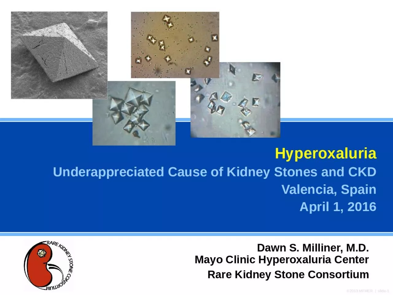
Dawn S. Milliner, M.D.Mayo Clinic Hyperoxaluria CenterRare Kidney Stone Consortium
HyperoxaluriaUnderappreciated Cause of Kidney Stones and CKDValencia, SpainApril 1, 2016
Slide254 y.o. woman with CKD stage 4
In good general health aside from marked obesity48 yrs of age gastric bypass followed by excellent weight loss over 2 years.51 y.o. seen for diarrhea. BMI 25.6. Stool fat 68 gm/24 hrs
(< 7 gm normal). Serum creatinine 1.6 mg/dl (< 1.1 normal). Bypass revised. 54 y.o. routine care for osteoporosis. S creatinine 3.9 mg/dl. Kidney size normal. Renal ultrasound increased echogenicity. Renal biopsy performed.
Slide354 y.o. woman with CKD stage 4
48 yrs of age marked obesity, gastric bypass followed by excellent weight loss over 2 years.51 y.o. seen for diarrhea. BMI 25.6. Stool fat 68 gm/24 hrs (< 7 gm
normal). Serum creatinine 1.6 mg/dl (<1.1 normal). Bypass revised. 54 y.o. routine care for osteoporosis. S creatinine 3.9 mg/dl. Kidney size normal. Renal ultrasound increased echogenicity. Renal biopsy performed.
Slide454 y.o. woman with CKD stage 4
55 y.o. preemptive LUD kidney transplant. Acute rejection treated with methylprednisolone. Creatinine remained 3.3 mg/dl, eGFR 15. CT showed 3 stones in allograft. Urine oxalate 0.92 mmol/24 hours (<0.46 normal).Low oxalate diet, calcium supplementation but CKD progressed. Second transplant planned.
Slide5Oxalate: Significance for the KidneySmall organic compound (C
2O4)Produced by the liver and ingested in the dietCannot be metabolized in humans, must be eliminated by kidney excretionWhen complexed with calcium is poorly soluble. Calcium oxalate crystals and kidney stones form in the urine.
Calcium oxalate crystals incite inflammation and tissue damageCKD and renal failure may result
Slide6Oxalate balance on a typical western diet
10%
30%
Diet
100 mg
Stool
9
0 mg
Glyoxylate
Ascorbic Acid
Endogenous Production
24 mg (1 mg/hr)
Absorbed
10 mg
Renal Excretion
34 mg
Slide7CP1285192-18
Aronson: KI, 2006
Ingested oxalate
Fecal oxalate
Urine oxalate
Plasma
oxalate
Hepatic
production
Intestinal
absorption
Intestinal
secretion
Normal
Oxalate Homeostasis
Slide8Hyperoxaluria Idiopathic Stone Formers
Enteric HyperabsorptionCrohn’s diseaseChronic pancreatitisCystic fibrosisSurgical resection of small bowelMalabsorptive bariatric surgery procedures
Medications (orlistat)Primary Hyperoxaluria
Slide9Urinary oxalate excretion in nonstone formers, routine stone formers, restrictive bariatric surgery subjects, and RYGB bariatric surgery subjects. Semins
MJ et al, J Urol 2010
Hyperoxaluria in Various Conditions
Slide10Enteric hyperoxaluria is caused by fat malabsorption
Ca
++
Ox
--
FA
BA
Ox
--
Slide11Lieske J: Kidney
Int
2015 Risk of new-onset nephrolithiasis after bariatric surgery. The risk of incident stones was greater after Roux-en-Y gastric bypass (RYGB) or malabsorptive bariatric procedures, compared with that in matched obese controls (P<0.001 overall). Patients with restrictive procedures were not at increased risk.
Nephrolithiasis after Bariatric Surgery
Slide12Inner medullary collecting duct (IMCD) deposits are mixture of apatite and calcium oxalate (CaOx). Two different large IMCD plugs from separate patients are seen under polarizing optics. These deposits show birefringent (arrowheads) and nonbirefringent (single arrows) crystals forming the same deposit.
Evan AP et al: Kidney Int 2010
Renal Histopathology in patients with small bowel
resection and calcium oxalate stone disease
Slide13Lieske J: Kidney
Int
2015 Risk of new-onset chronic kidney disease (CKD) after bariatric surgery. The risk of incident CKD was greater after malabsorptive bariatric procedures compared with that in matched obese controls (P=0.004 overall).
CKD after Bariatric Surgery
Slide14Trends in the Numbers of Bariatric Surgery Procedures Worldwide: 2003
to 2011
Buchwald H,
Obes Surg 2013
Slide15Idiopathic stone disease
Frequency 8-12% of populationCaOx stones 75-80%Hyperoxaluria 15-20%
Urolithiasis and the Risk of ESRDConclusions: Symptomatic stone formers are at increased risk for ESRDindependent of several cardiovascular risk factors. Other urologicaldisease is relatively common among stone formers who develop ESRD.
Clin J Am Soc Nephrol 7:1409-1415, 2012.
Slide16El-Zoghby Z: CJASN 2012
Incidence of Renal Failure in Stone Formers
versus Age-matched Controlsp = 0.01
Slide17Primary Hyperoxaluria
A
Model for Oxalate Nephropathy
Inherited inborn error of metabolism with marked hepatic overproduction of oxalate
3 types described due to deficiencies of hepatic AGT (PH1), GRPHR (PH2), or HOGA (PH3)Urine oxalate 2-8 x normal from birthStones,
nephrocalcinosis, and loss of kidney function over time are characteristic Among patients with PH1 75% have ESKD by 50
yrs of age
Slide18Primary Hyperoxaluria
Histopathologic Examination of
Kidney Tissue
Calcium oxalate crystals in proximal proximal
tubule cells of PH type 1 patients with preserved kidney function
Worcester et al, AJP 2013
Oxalate concentration highest in S3 segment of proximal
tubule
Proximal tubule
fluid is supersaturated for
CaOx
in PH1
Slide19Rare Kidney Stone Consortium
Primary Hyperoxaluria Registry, 387 Patients
73%
9%
9
%
7
%
Slide20The Primary Hyperoxalurias
Slide21Patients (no.)
Primary Hyperoxaluria Kidney Status at Diagnosis
Age0-4
5-910-14
15-1920-24
25-2930-34
35-3940-44
45-49
50-54
55+
Preserved renal function
End stage renal failure
Slide22Renal Survival in Primary Hyperoxaluria
Renal Survival plots showing poorer renal survival for PH1 patients followed by PH2. Kaplan-Meier renal survival plot of the PH1, PH2, PH3, and NMD cohorts. Tables below Kaplan-Meier plots show survival estimates with number of patients at risk in parentheses.
at risk in parentheses.
Slide23Primary Hyperoxaluria15
y.o. boy with his second kidney stone28 y.o. with ESKD and dense kidneys on imaging14 m.o. with failure to thrive found to have stage 4 CKD61 y.o. undergoing transplant evaluation for ESKD of unknown etiology (single stone on imaging)
43 y.o. for stone evaluation with normal kidney function
Slide24Diagnosis of the Primary Hyperoxalurias
CP1304636-1
or
Renal insufficiency
Ca oxalate stones
Nephrocalcinosis
Ca oxalate tissue deposits
Uox
>0.7 mmol/1.73 m
2
/24 hr
or
Uox
/
ucreat
> normal for age
Uox
>0.5 mmol/1.73 m
2
/24 hr
and/or
Plasma ox > 20
mol/L
Evaluate
Secondary cause?
No
DNA testing
Yes
Normal renal function
Caox
stones or nephrocalcinosis in childhood
Recurrent
CaOx
stones/ hyperoxaluria in adults
Hyperoxaluria with family
hx
of PH
Slide25Mulay
SR.
Nephrol Dial Transplant 2013Routes and Sites of Crystal Deposition in the Kidney
Slide26Hyperoxaluria: Mechanisms of injury
Caox crystals can trigger injury when deposited within the kidney. Mechanisms largely unknown.Intracellular NLRP3 inflammasome discovered: a pattern recognition platform that translates crystal uptake into immune activation through secretin of IL-1B and IL-18. Proof of concept in animal models of oxalate nephropathy (
Knauf KI 2013).Crystal-induced Inflammatory infiltrate, multinucleated giant cells in patients with hyperoxaluria.Scarring, nephrocalcinosis
Slide27Mulay
SR.
Nephrol Dial Transplant 2013Mechanisms of Crystal-Induced Renal Inflammation
Slide28Oxalate Nephropathy Awareness with early and specific diagnosis
Targeted intervention for primary or secondary cause, as indicated. Reduce oxalate produced (e.g. pyridoxine in PH) or absorbed (enteric hyperoxaluria)Reduce calcium oxalate crystal formation, deposition
Future directions anti-inflammatory strategiesParticular attention to renal replacement therapies.
Slide29Acknowledgements
Rare Kidney Stone Consortium
Staff
RKSC Coordinating Centers
T
he
many
contributors to the PH Registry