
It has a sensory function The regions of the dorsal horns the nuclei of the dorsal gray column are sensory in nature and are arranged in four groups from dorsal to ventral 1Dorsomarginal nucleus extends the entire length of the spinal cord capping the dorsal horn and receives afferent fib ID: 914837
Download Presentation The PPT/PDF document "Posterior (dorsal) horn:" is the property of its rightful owner. Permission is granted to download and print the materials on this web site for personal, non-commercial use only, and to display it on your personal computer provided you do not modify the materials and that you retain all copyright notices contained in the materials. By downloading content from our website, you accept the terms of this agreement.

![expressiveimpactofnominalschemasingeneral[5].Beyondthissingularresult,](https://thumbs.docslides.com/484247/expressiveimpactofnominalschemasingeneral-5-beyondthissingu.jpg)

Slide1
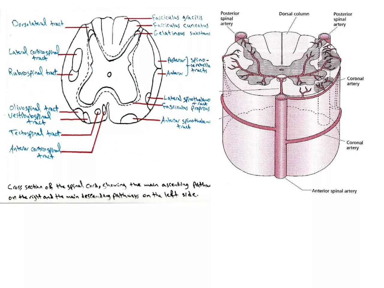
Slide2Posterior (dorsal) horn: It has a sensory function.The regions of the dorsal horns:
the nuclei of the dorsal gray column are sensory in nature and are arranged in four groups from dorsal to ventral:
1-Dorsomarginal nucleus: extends the entire length of the spinal cord, capping the dorsal horn, and receives afferent fibers carrying pain, temperature and light touch sensation.2-Substantia gelatinosa of Rolando: It is situated next to dorsomarginal group of nuclei and it presents throughout the length of the spinal cord. It acts as an editor of the sensory input (modifying the arriving pain and temperature sensation).3-Nucleus proprius (it is the main sensory nucleus): It receives nerve fibers from the substantia gelatinosa. It also presents throughout the length of the spinal cord. Nucleus proprius receives pain, temperature and light touch sensations and provides input to the lateral and ventral (anterior) spinothalamic tracts. 4-Nucleus dorsalis of Clark: It is situated near the base of the dorsal horn and it presents only between (C8 and L3). It concerns with the posterior spinocerebellar tract for proprioceptive sensation.
Slide3Intermediate horn: In the thoracic region, it is responsible for sympathetic output (between T1 and L2 segment). While in the sacral region, it is responsible for parasympathetic output (S2-S4 segment).
Slide4White matter
of the spinal cord is outside the gray matter and it divides into: dorsal column, anterior column and lateral column.The white matter of the spinal cord contains two major types of nerve fibers:Descending tracts (motor) from high centers of the brain.
Ascending tracts (sensory) to high centers of the brain.
Slide5Descending tracts:
Corticospinal tract (pyramidal tract).
Rubrospinal tract.
Vestibulospinal tract. Reticulospinal tract. Tectospinal tract.
The rubrospinal, reticulospinal, tectospinal and vestibulospinal tracts are parts of the extrapyramidal tracts or system.
Slide6Corticospinal tract:
It is concerning with the initiation of voluntary movement. This tract begins from the motor area of the cerebral cortex and then down till the medulla oblongata. The majority of these fibers cross to other side in the medulla oblongata in the decussation region that called the pyramid. These crossed fibers are called the
lateral corticospinal tract. The rest of the fibers are called the
anterior corticospinal tract that crossed in the spinal cord. Thus, the majority of the corticospinal tract is crossed to other side, either as lateral corticospinal tract (90%), or as anterior corticospinal tract (8%), but these two tracts are differed in the level of decussation. 2% of corticospinal tract is uncrossed.
Slide7Slide8Slide92- Rubrospinal tract:
It is an alternative pathway of the corticospinal tract and it is responsible for gain or return of the motor activity after damage of the corticospinal tract.
The rubrospinal tract starts from the motor area to red nucleus of the midbrain and then crosses to other side and forming the rubrospinal tract.3- Tectospinal tract:
It starts from the superior colliculus of the tectum of the midbrain and carry information from the eyes to the spinal tract through the tectospinal tract. It is crossed fibers and its function is to adjust the posture according to the visual stimuli.4- Vestibulospinal tract:
It is uncrossed tract from the lateral vestibular nucleus to the spinal cord. It is important for maintaining the equilibrium of the posture.5- Reticulospinal tract:
They are of two types:
Slide10Pontine reticulospinal tract: these are uncrossed fibers from the reticular formation of the pons.
Medullary reticulospinal tract: these fibers contain both crossed and uncrossed fibers.
The function of the reticulospinal tract is for the control of the posture unconsciously.
Slide11Slide12Ascending tracts (sensory):
1- The pathway for pain and temperature is the
lateral spinothalamic tract. The first order neuron is the neuron in the dorsal root ganglia → nucleus proprius of the dorsal horn of the spinal cord → fibers cross to other side to form the lateral spinothalamic tract → thalamus (3
rd order neuron) → sensory cortex of the cerebral hemisphere.2- The pathway for light touch and pressure is the anterior spinothalamic tract.
The first order neuron is in the dorsal root ganglia → nucleus proprius of dorsal horn of the spinal cord → cross the midline → anterior spinothalamic tract → thalamus → sensory cortex of the cerebral hemisphere.
Slide133- Pathway for discriminative touch (two points discrimination) (
fasciculus
gracilis) and conscious proprioception (fasciculus cuneatus). The proprioception is the sense of position. The first order neuron is in the dorsal root ganglia → nucleus
gracilis or cuneatus of the medulla oblongata → cross to other side → forming the medial lemniscus → ascend to the thalamus (ventro-posterior nucleus of the thalamus) → cerebral cortex.4- Pathway for unconscious proprioception: it is throughAnterior (ventral) spinocerebellar tract.
Posterior (dorsal) spinocerebellar tract.Cuneocerebellar tract.
Slide14A- Anterior spinocerebellar tract:
Dorsal root ganglia (first order neuron) → spinal cord (2
nd order neuron) → go to the same side of the cerebellum.B- Posterior spinocerebellar tract: The first order neuron in the dorsal root ganglia → 2nd
order neuron in the nucleus dorsalis of Clarke → same side of the cerebellum (no cross).C- Cuneocerebellar tract: It is a small tract, start from the accessory cuneate nucleus of the medulla oblongata, and pass through the brain stem, then to the cerebellum (of the same side). If these unconscious proproceptive tracts damage → ataxia
.
Slide15Intersegmental tracts:
These tracts communicate the spinal cord parts together:
Dorso-lateral tract that lie between tip of the posterior horn and surface of the spinal cord.
Fasciculus proprius which is around the gray matter of the spinal cord.