/
Lecture #9 First semester Lecture #9 First semester

Lecture #9 First semester - PowerPoint Presentation

candy
candy . @candy
Follow
70 views
Uploaded On 2024-01-03

Lecture #9 First semester - PPT Presentation

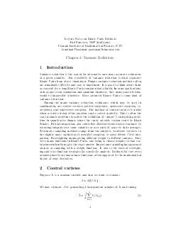
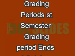
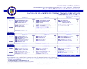
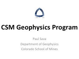
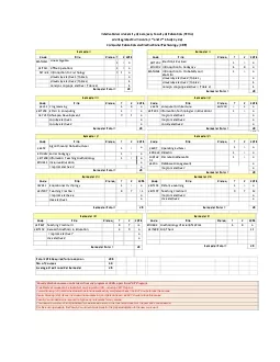
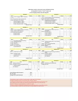
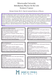
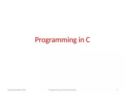
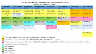
Cardiovascular D isorders I Hypertension Al Mustaqbal University College College of Nursing 2 nd Class Adult Nursing by lecturers DrFakhria Jaber Definition ID: 1037685

artery blood pressure coronary blood artery coronary pressure oxygen heart pain hypertension myocardial increased flow cardiac nursing disease ischemia

Share:

Link:

Embed:

Download Presentation from below link

Download Presentation The PPT/PDF document "Lecture #9 First semester" is the property of its rightful owner. Permission is granted to download and print the materials on this web site for personal, non-commercial use only, and to display it on your personal computer provided you do not modify the materials and that you retain all copyright notices contained in the materials. By downloading content from our website, you accept the terms of this agreement.


Presentation Transcript

1. Lecture #9First semester Cardiovascular DisordersI- HypertensionAl-Mustaqbal University CollegeCollege of Nursing 2nd ClassAdult Nursing :bylecturers Dr.Fakhria Jaber

2. DefinitionHypertension is defined by the American Society of Hypertension (ASH) and the International Society of Hypertension (ISH) as: a systolic blood pressure (SBP) of 140 mm Hg or higher or a diastolic blood pressure (DBP) of 90 mm Hg or higher, based on the average of two or more accurate blood pressure measurements taken 1 to 4 weeks apart by a health care provider.

3. Thanks For Listening

4. PathophysiologyEach time the heart contracts, pressure is transferred fro the contraction of the heart muscle to the blood and then pressure is exerted by the blood as it flows through the blood vessels.

5. Causes of hypertensionAbout 95% of patients with high blood pressure have primary hypertension (also called essential hypertension), which is defined as high blood pressure from an unidentified cause. The remaining small percentage, about 5%, have secondary hypertension, which occurs when a cause for the high blood pressure can be identified.These causes include chronic kidney disease, hyperaldosteronism (mineralocorticoid hypertension). High blood pressure can also occur with pregnancy;

6. Prolonged blood pressure elevation eventually damages blood vessels throughout the body, particularly in target organs such as the heart, kidneys, brain, and eyes.The usual consequences of prolonged, uncontrolled hypertension are myocardial infarction, heart failure, renal failure, strokes, and impaired vision.The left ventricle of the heart may become enlarged (left ventricular hypertrophy) as it works to pump blood against the elevated pressure Cont.…

7. Hypertension signs and symptoms

8.

9.

10.

11. Treatment

12. Monitor BPObtain complete historyto assess for symptoms that indicate target organ damage (whether other body systems have been affected by the elevated blood pressure).Ex: anginal pain; shortness of breath; alterations in speech, vision, or balance; nosebleeds; headaches; dizziness; or nocturia.Pulserate, rhythm, and character of apical and peripheral pulsesAssessment

13. Deficient knowledge regarding the relation between the treatment regimen and control of the disease process.Noncompliance with therapeutic regimen related to side effects of prescribed therapy.

14. objective : lowering and controlling the blood pressure without adverse effects.support and teach the patient to adhere to treatment regimen.Implement necessary lifestyle changesTake medications as prescribedSchedule regular follow-up appointmentsTeach disease process and how lifestyle changes and meds can control hypertension.emphasize concept of controlling hypertension rather than curing it

15. II. Coronary artery diseases1- Angina pectoris

16. Coronary AtherosclerosisCoronary artery disease (CAD) is the most prevalent type of cardiovascular disease in adults. The most common cause of coronary artery disease (CAD) in the world is: Atherosclerosis, an abnormal accumulation of lipid, or fatty substances, and fibrous tissue in the lining of arterial blood vessel walls. These substances block and narrow the coronary vessels in a way that reduces blood flow to the myocardiumAtherosclerosis involves a repetitious inflammatory response to injury of the artery wall and subsequent alteration in the structural and biochemical properties of the arterial walls.

17.

18.

19. I: Angina pectorisAngina pectoris is a clinical syndrome usually characterizedby episodes of pain or pressure in the anterior chest.The cause is usually insufficient coronary blood flow.The insufficient flow results in a decreased oxygen supply to meet an increased myocardial demand for oxygen in response to physical exertion or emotional stress.In other words, the need for oxygen exceeds the supply.

20. Angina is usually caused by atherosclerotic disease.Almost invariably, angina is associated with a significant obstruction of at least one major coronary artery. Normally, the myocardium extracts a large amount of oxygen from the coronary circulation to meet its continuous demands.When demand increases, flow through the coronary arteries needs to be increased. When there is a blockage in a coronary artery, flow cannot be increased and ischemia results.Pathophysiology

21. Factors are associated with anginal pain:Physical exertion, which precipitates an attack by increasing myocardial oxygen demandExposure to cold, which causes vasoconstriction and elevated blood pressure, with increased oxygen demandEating a heavy meal, which increases the blood flow to the mesenteric area for digestion, thereby reducing the blood supply available to the heart muscle; in a severely compromised heart, hunting of blood for digestion can be sufficient to induce anginal painStress or any emotion-provoking situation, causing the release of catecholamines, which increases blood pressure, heart rate, and myocardial workload

22.

23. Ischemia of the heart muscle may produce pain or other symptoms, varying from mild indigestion to a choking or heavy sensation in the upper chest. The pain may be accompanied by severe apprehension and a feeling of impending death. It is often felt deep in the chest behind the sternum (retrosternal area). Typically, the pain or discomfort is poorly localized and may radiate to the neck, jaw, shoulders, and inner aspects of the upper arms, usually the left arm. Clinical Manifestations

24. The diagnosis of angina begins with the patient’s history related to the clinical manifestations of ischemia. A 12-lead electrocardiogram (ECG) may show changes indicative of ischemia, such as T-wave inversion, ST segment elevation. Laboratory studies are performed; these generally include cardiac biomarker testing to rule out (Myocardial Infarction section). The patient may undergo an exercise or pharmacologic stress test in which the heart is monitored continuously by an ECG, echocardiogram, or both. The patient may also be referred for a nuclear scan or invasive procedure (e.g., cardiac catheterization, coronary angiography).Assessment and Diagnostic Findings

25. Medical ManagementThe objectives of the medical management of angina are to decrease the oxygen demand of the myocardium and to increase the oxygen supply. Medically, these objectives are met through pharmacologic therapy and control of risk factors.Alternatively, reperfusion procedures may be used to restore the blood supply to the myocardium. These include PCI procedures (e.g., percutaneous transluminal coronary angioplasty [PTCA] and intracoronary stents) and (CABG) Coronary artery bypass graft.

26. Pharmacologic therapy

27. NURSING ASSESSMENT

28.

29. Encourage to stop all activities and sit or rest in bed in a semi-fowler’s positionMeasuring Vital SignsObserve for signs of respiratory distressAdminister Nitroglycerin as prescribedAdminister Oxygen therapy if the patient’s respiratory rate is increased or oxygen saturation is decreasedNURSING INTERVENTIONS

30. Coronary artery diseasesII: Myocardial infarctionMI refers to the process by which areas of myocardial cells in the heart are permanently destroyed.MI is usually caused by reduced blood flow in a coronary artery due to atherosclerosis and occlusion of an artery by an embolus or thrombus.Other causes of an MI include vasospasm (sudden constriction or narrowing) of a coronary artery; decreased oxygen supply (eg, from acute blood loss, anemia, or low blood pressure); and increased demand for oxygen (eg, from a rapid heart rate, thyrotoxicosis, or ingestion of cocaine).

31. 1. CARDIOVASCULAR• Chest pain : chest pain occurs suddenly, severe immobilizing chest painthat not relieved by rest ,position change, and medications.• Increased jugular venous distention• BP may be elevated because of sympathetic stimulation or decreased BPbecause of decreased contractility, development if cardiogenic shock• Decrease pulse rate• ST- segment and T-wave changes, ECG may show tachycardia,bradycardia, or dysrhythmias.2. Respiratory• Shortness of breath (SOB)• Dyspnea, tachypnea, and crackles if MI hascaused pulmonary congestion.• Pulmonary edemaClinical manifestations of MI

32. 3. Gastrointestinal or GIT• Nausea and vomiting.4. Genitourinary• Decreased urinary output may indicatecariogenic Shock.5. Skin• Cool , clammy, diaphoretic, and paleappearance on skin.6. Neurologic symptoms• Anxiety, restlessness,and light headness.7. Psychological• Fear with feeling of impending doom orpatient may deny that anything is worng.Cont..

33. ECG Changes in MIEffects of ischemia, injury, and infarction on ECG recording. Ischemia causes inversion of T wave because of altered repolarization.Cardiac muscle injury causes elevation of the ST segmentQ waves develop because of the absence of depolarization current from the necrotic tissue and opposing currents from other parts of the heart.

34. • Laboratory tests called “CARDIAC BIOMARKERS” are used to diagnose MI.• Creatine kinase –MB or CK-MB• myoglobin• Troponin T or ILaboratory tests

35. Pharmacologic therapy for MI: The patient with an acute MI receives the same medications as the patient with unstable angina, with the possible additions ofThrombolytics: Streptokinase( Kabikinase, Streptase) Alteplase (Activase ) , Reteplase, Anistreplase (Eminase)Analgesics: morphine sulfate (Duramorph, Astramorph)Angiotensin-converting enzyme (ACE) inhibitors.Patients should receive a beta-blocker initially

36. Invasive Coronary Artery ProceduresPTCA is carried out in the cardiac catheterization laboratory. Hollow catheters called sheaths are inserted, usually in the femoral artery (and sometimes the radial artery), providing a conduit for other catheters.Catheters are then threaded through the femoral artery, up through the aorta, and into the coronary arteries. Angiography is performed using injected radiopaque contrast agents (commonly called dye) to identify the location and extent of the blockage.

37. A balloon-tipped dilation catheter is passed through the sheath and positioned over the lesion. The physician determines the catheter position by examining markers on the balloon that can be seen with fluoroscopy.When the catheter is properly positioned, the balloon is inflated with high pressure for several seconds and then deflated. The pressure compresses and often “cracks” the atheroma. The media and adventitia of the coronary artery are also stretched.

38. Coronary Artery StentAfter PTCA, a portion of the plaque that was not removed may block the artery. The coronary artery may recoil (constrict) and the tissue remodels, increasing the risk for restenosis.A coronary artery stent is placed to overcome these risks. A stent is a woven mesh that provides structural support to a vessel at risk of acute closure. The stent is placed over the angioplasty balloon.When the balloon is inflated, the mesh expands and presses against the vessel wall, holding the artery open.

39. The balloon is withdrawn, but the stent is left permanently in place within the artery.Eventually, endothelium covers the stent and it is incorporated into the vessel wall.Because of the risk of thrombus formation in the stent, the patient receives antiplatelet medication eg, clopidogrel and lifetime use of aspirin.

40. Surgical proceduresCoronary Artery RevascularizationCABG is a surgical procedure in which a blood vessel from another part of the body is grafted to the occluded coronary artery so that blood can flow beyond the occlusion; it is also called a bypass graft.The right and left internal mammary arteries and, occasionally, radial arteries are also used for CABG.Arterial grafts are preferred to vein grafts because they do not develop atherosclerotic changes as quickly and remain patent longer.

41. The vessel most commonly used for CABG is the greater saphenous vein, followed by the lesser saphenous vein Cephalic and basilic veins are used also.The vein is removed from the leg (or arm) and grafted to the ascending aorta and to the coronary artery distal to the lesion

42. NURSING AssessmentOne of the most important aspects of care of the patient with MI is the assessment. It establishes the patient’s baseline, identifies the patient’s needs, and helps determine the priority of those needs. Systematic assessment includes a careful history, particularly as it relates to symptoms: chest pain or discomfort, dyspnea (difficulty breathing), palpitations, unusual fatigue, syncope (faintness), or other possible indicators of myocardial ischemia. Each symptom must be evaluated with regard to time, duration, and the factors that precipitate the symptom and relieve it, and in comparison with previous symptoms.

43. NURSING DIAGNOSESBased on the clinical manifestations, history, and diagnostic assessv c ment data, major nursing diagnoses may include:Acute pain associated with increased myocardial oxygen demand and decreased myocardial oxygen supplyRisk for impaired cardiac function associated with reduced coronary blood flowRisk for hypovolaemiaImpaired peripheral tissue perfusion associated with impaired cardiac output from left ventricular dysfunctionAnxiety associated with cardiac event and possible deathLack of knowledge about post-ACS self-care

44. Nursing InterventionsRELIEVING PAIN AND OTHER SIGNS AND SYMPTOMS OF ISCHEMIAIMPROVING RESPIRATORY FUNCTIONPROMOTING ADEQUATE TISSUE PERFUSIONREDUCING ANXIETY