
6619 European Review for Medical and Pharmacological Sciences2021 25 66196622 V BAGNARA M CASTAGNETTI AE CALOGERO RA CONDORELLIR CANNARELLA 6620 eration was performed under general anesth ID: 958780
Download Pdf The PPT/PDF document "Abstract OBJECTIVE The leiomyoma is a be..." is the property of its rightful owner. Permission is granted to download and print the materials on this web site for personal, non-commercial use only, and to display it on your personal computer provided you do not modify the materials and that you retain all copyright notices contained in the materials. By downloading content from our website, you accept the terms of this agreement.


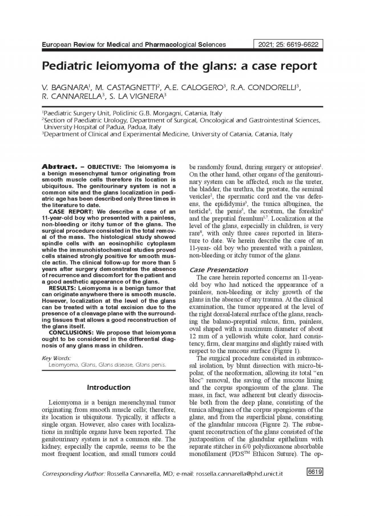
6619 Abstract. OBJECTIVE: The leiomyoma is a benign mesenchymal tumor originating from smooth muscle cells therefore its location is ubiquitous. The genitourinary system is not a common site and the glans localization in pediatric age has been described only three times in the literature to date. CASE REPORT: We describe a case of an 11-year-old boy who presented with a painless, non-bleeding or itchy tumor of the glans. The surgical procedure consisted in the total remov European Review for Medical and Pharmacological Sciences2021; 25: 6619-6622 V. BAGNARA, M. CASTAGNETTI, A.E. CALOGERO, R.A. CONDORELLIR. CANNARELLA 6620 eration was performed under general anesthesia with penile block, without the use of tourniquets or adrenaline solutions. Only for better patient’s postoperative comfort, a 12 Ch bladder catheter was kept in place for 24 hours.The histopathological study described a well-circumscribed lesion due to the presence of a pseudocapsule of homogeneous appearance and consisting of fusocellular elements with eosinophilic cytoplasm, spindle nucleus with rounded edges. On immunohistochemical examination, the cellular structure showed high cytoplasmic positivity to speci�c smooth muscle alpha-actin and desmin while the S-100 protein resulted negative (Figure 3). The absence of cellular atypia, areas of necrosis and rare or absent mitosis �nally allowed to exclude malignant nature of the lesion. The clinical follow-up for more than �ve years after surgery demonstrates the absence of recurrence and discomfort for the patient and a good aesthetic appearance of the glans (Figure 4).DiscussionLeiomyoma is a benign tumor that can originate anywhere there is smooth muscle. The origi
n of glans leiomyoma could be explained by the presence of smooth muscle cells in the blood vessel wall and in the trabeculae that separate the vascular lacunae of the corpus spongiosumHowever, localization at the level of the glans is very rare and only a few cases have been described in the literature to date. A systemic review of the medical literature using the keywords “leiomyoma”, “glans”, “glans disease” and “glans penis” showed only 3 cases of pediatric glans leiomyoma reported up to June 2021. The �rst case of glans leiomyoma was reported by Belis in 1979 and concerned a 3-year-old child. Since then, only two other cases have been described by Stehr et al in 2000, in a 12-year-old teenager, and by Redman in 2000 in an 8-year-old boy11(Table I). However, in the case reported by Stehret al the excision of leiomyoma was only partial but the follow-up at 18 months showed the absence of symptoms. Other sporadic cases of glans leiomyoma have been described in adults.Figure 1.Clinical appearance of the lesion. Figure 2.Appearance of the tumor after glans mucosa detachment (); Appearance of the glans afterthe “en bloc” excision of the tumor (); Tumor after excision ( 6621 In all cases of glans’ localization, the treatment consisted in a total or partial excision of the tumor.In no case a recurrence or malignant transformation is reported. In our experience, the total “en bloc” exeresis of the neoformation was possible by the presence of a cleavage plane that allowed an accurate and complete dissection of the lesion, saving the mucosa and the erectile tissue, corpus spongiosum of the glans, both adherent but not affected by the lesion and, ther
efore, dissociable. The saving healthy tissue is certainly necessary for a more accurate reconstruction of the glans and limit serious and signi�cant impairments both aesthetically and functionally.In fact, after one year, the glans depression, residual on removal, and the surgical scar were barely visible. Furthermore, the boy did not suffer from discomfort, such as pain and/or itching, nor loss of sensitivity. ConclusionsAlthough leiomyoma localized in the glans is a very rare condition, it must be kept in mind in the differential diagnosis of tumors that can occur here, such as plexiform neuro�broma, smooth muscle cell hamartoma, schwannoma, nerve sheath myoma and �brohistiocytoma11,12The early and complete exeresis of this tumor is necessary to clarify the diagnosis and prevent further growth. In fact, even if con�ned to the subcutaneous layer, the growth of this tumor could lead to further dif�culties in the excision and subsequent reconstruction of the glans with possible major repercussions both on an aesthetic and on a functional point of view.Figure 3.(H&E stain) Spindle cells with an eosinophilic cytoplasm at low () and high magni�cation (); Immunohistochemical staining for smooth muscle actin ( Figure 4. Clinical appearance of the glans penis at one year follow-up. AuthorYear of publicationPatients’ age (yr)LocationDimensions (cm)Local recurrenceBelis et al1979Glans penis1.5NoStehr et al200012Glans penisNoRedman et al2000Glans penisNoOur case202111Glans penis1.2No Pediatric leiomyoma of the glans: a case report 6622 Conflict of InterestThe Authors declare that they have no con�ict of interests.Informed Consent StatementInforme
d consent was obtained from all subjects involved in the study.References1)Belis JA, Post GJ, Rochman SC, Milam DF. Genitourinary leiomyomas. Urology 1979; 13: 424-429.2)Mendrek M, Bach C, Gaisa NT, Vögeli TA. Leiomyoma arising from the right seminal duct/seminal vesicle—Report of a rare case and review of the literature. Andrologia 2019; 51: e13174.3)Ozden O, Orhan D, Karnak I. Epididymal leiomyoma: an unusual intrascrotal tumor in a child. J Pediatr Surg 2009; 44: e5-7.4)Kullolli VS, Kullolli S, Pawar S, Gautam D. Leiomyoma of testis -a case report. Indian J Surg 2011; 73: 233-235.5)Liu SP, Shun CT, Chang SJ, Chen J, Hsieh JT. Leiomyoma of the corpus cavernosum of the penis. Int J Urol 2007; 14: 257-258.6)Leoni S, Prandi S, Mora A. Leiomyoma of the prepuce. Eur Urol 1980; 6: 188-189.7)Moch H, Cubilla AL, Humphrey PA, Reuter VE, Ulbright TM. The 2016 WHO Classi�cation of Tumours of the Urinary System and Male Genital Organs-Part A: Renal, Penile, and Testicular Tumours. Eur Urol 2016; 70: 93-105. 8)Dehner LP, Smith BH. Soft tissue tumors of the penis. A clinicopathologic study of 46 cases. Cancer 1970; 25: 1431-1447.9)Christ GJ. The penis as a vascular organ. The importance of corporal smooth muscle tone in the control of erection. Urol Clin North Am 1995; 22: 727-745. 10)Stehr M, Rohrbach H, Schuster T, Dietz HG. Leiomyom der Glans Penis [Leiomyoma of the glans penis]. Urologe A 2000; 39: 171-173.11)Redman JF, Liang X, Ferguson MA, Savell VH. Leiomyoma of the glans penis in a child. J Urol 2000; 164: 791.12)Bartoletti R, Gacci M, Nesi G, Franchi A, Rizzo M. Leiomyoma of the corona glans penis. Urology 2002; 59: 445. V. Bagnara, M. Castagnetti, A.E. Calogero, R.A. Condorelli, R. Cannarella, S. La Vign