
1 Protein Chains are Polymers of Amino Acids 2 Isoelectric Point 3 NH 3 CH 2 CO 2 H H 2 O NH 3 CH 2 CO 2 H 3 O pKa 234 NH 3 CH 2 ID: 927470
Download Presentation The PPT/PDF document "Protein Structure Lecture 5" is the property of its rightful owner. Permission is granted to download and print the materials on this web site for personal, non-commercial use only, and to display it on your personal computer provided you do not modify the materials and that you retain all copyright notices contained in the materials. By downloading content from our website, you accept the terms of this agreement.


Slide1
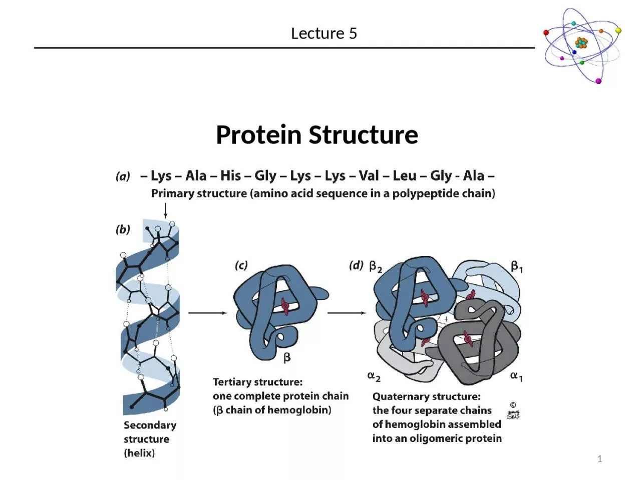
Protein Structure
Lecture 5
1
Slide2Protein Chains are Polymers of Amino Acids
2
Slide3Isoelectric Point
3
(NH
3
CH
2
CO
2
H)
+
+
H
2O ⇌
(NH3CH2CO2)
+ H3O+ pKa
= 2.34(NH3CH2
CO2) + H2O
⇌ (NH2CH2CO2
)- + H3O
+ pKa = 9.69
The pH at which a zwitterion carries no
net charge
C
pK
a,1
pK
a,1
Expected pH @ equivalence for HA with
pKa = 2.34
Actual pH @ equivalence for glycine
pK
a,2
Slide4Isoelectric Point
4
(NH
3
CH
2
CO
2
H)
+
+
H
2O ⇌
(NH3CH2CO2)
+ H3O+ pKa
= 2.34(NH3CH2
CO2) + H2O
⇌ (NH2CH2CO2
)- + H3O
+ pKa = 9.69
The pH at which a zwitterion carries no
net charge
Slide5Isoelectric Point
5
AH
3
2+
+
H
2
O
⇌
AH
2
+
+ H3
O+ pKa = 2.34AH2
+ + H2O ⇌ AH
+ H3O+
pKa = 6.04AH +
H2O ⇌ A- +
H3O+ pKa =
9.69
The pH at which a zwitterion carries no net charge
Slide6Common Side Chain Chemistry
6
Disulfide Formation
Phosphorylation of Side Chain
Slide7The Peptide Bond
7
A
B
C
D
Bond
Expected
Bond Length (Å)
Actual
Bond Length (Å)
A
1.46
1.33
B
1.51
1.51
C
1.22
1.24
D
1.46
1.46
**
**
What can explain the deviation from expected values?
Slide8The Peptide Bond
8
~60%
~40%
So the peptide bond is planar
c
is
and trans conformations can be envisioned
Slide9The Peptide Bond
9
Slide10The Peptide Bond
10
Polypeptide chains are sequences of peptide bond planes joining at every
C
a
Modest rotation of one plane relative to another will dramatically influence the stability of a polypeptide
Steric restrictions will disallow this conformation
Slide11Torsion Angles
11
Slide12Ramachandran
Plot
12
There are only a few “allowable” combinations of
f
and
y
.
Notable exceptions:
The side chain of
Proline
restricts
f
≈ -60°
Glycine does not have a
b carbon and can assume
f,y combinations that are forbidden in other amino acids
All other regions of the
ramachandran
plot are
sterically forbidden
Slide13Ramachandran
Plot
13
For full length proteins, several areas appear
b
sheets
Centered around
F
= -139,
Y
= 135
Helical structures
Centered around
F
= -57,
Y
= 47
Slide14b
Sheets
14
b
sheets occur when two nearly fully extended (
F
= -139,
Y
= 135
) polypeptide chains interact
Significant hydrogen-bonding between O and N of opposite chains
Because the chains are extended, the sheets take on a pleated appearance
N-term
C
-term
Parallel
Antiparallel
Note the alternating orientation of the side chains
Slide15Parallel
b Sheets
15
C
Term
C
Term
N Term
N Term
Slide16Antiparallel
b Sheets
16
N Term
N Term
C
Term
C
Term
Slide17Helices
17
Helices occur when one peptide chain coils up (
F
= -57,
Y
=
47)
Significant hydrogen-bonding between backbone carbonyl Oxygen and amide Nitrogen
d
+
d
+
Helices are polar
Note the side chains decorate the exterior of the helix
Slide18Other 2˚ Structural Motifs
18
Random Coil
When the peptide backbone adopts no repeating structural features. It is not correct to say the protein is disordered in random coil.
Turns
Abrupt changes in direction in polypeptide structures.
Type
Separating Residues
d
1
g
2
b
3
a
4
P
5
F
2
=
-60, Y
2 = -30F3
= -90, Y3 = 0
F
2 = -60, Y2 = 120
F3 = -90,
Y3 = 0
Slide19Fibrous Proteins vs. Globular Proteins
19
Not Repeating
Repeating Primary Structures
Keratin – Form
Coiled Coils
Heptad Repeat
a b c d e f g
Tropomyosin
PDBid
1IC2
Slide20Tertiary Structure
20
Secondary Structural elements send to form so that Polar side chains and non-Polar side chains form opposite ‘faces’
Non-Polar
Polar
Backbone
Hydrophobic Regions pack together to form the core of globular proteins
Slide21Tertiary Structure
21
Structural Motifs (or Super Secondary Structure)
Certain Combinations of secondary structural elements tend to be found very commonly in protein structures
Slide22Tertiary Structure
22
Proteins are represented by showing how 2
nd
structures interact with each other.
Arrow are
b
sheets
Coils are
a
helices