
Placental MSC Exosomes and the Lessons Learned in the Last 2 Years Douglas J Spiel MD Stem Cells How do they work Totipotential Stem Cells Pluripotential Stem Cells Multipotential Stem Cells ID: 1010777
Download Presentation The PPT/PDF document "Central Nervous System Injuries in Child..." is the property of its rightful owner. Permission is granted to download and print the materials on this web site for personal, non-commercial use only, and to display it on your personal computer provided you do not modify the materials and that you retain all copyright notices contained in the materials. By downloading content from our website, you accept the terms of this agreement.


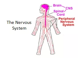


1. Central Nervous System Injuries in ChildrenPlacental MSC Exosomes and the Lessons Learned in the Last 2 YearsDouglas J. Spiel, MD
2. Stem Cells How do they work?Totipotential Stem CellsPluripotential Stem CellsMultipotential Stem CellsResident Progenitor Cells
3. Autologous vs Allogeneic Cells
4. Arnold Caplan:2006 – “The trophic effects of MSCs may have profound clinical use.”2017 – “I now urge that we change the name of MSCs to Medicinal Signaling Cells …”“It is, indeed, the patient’s own site-specific and tissue specific resident stem cells that construct the new tissue as stimulated by the bioactive factors secreted by the exogenously supplied MSCs” Revisionist Viewpoint!MSC Exosomes
5. The origin of biohacking dates back 150 odd years to physiologist Paul Bert in 1864 - ParabiosisBy splicing the blood circulation of 2 animals together we have shown that young blood rejuvenates old tissues, bones, muscles, brain, and nerve tissue.Nik Spencer/Nature; Chart Data: A. Eggel & T. Wyss-Coray Swiss Med. Wkly 144, W13914 (2014)[Photograph]. Retrieved from https://www.nature.com/news/ageing-research-blood-to-blood-1.16762What can Science teach us regarding aging?
6. Why does plasma transfer work?Are we transfusing stem cells? No. Simply, there are not enough stem cells in blood/plasma. In fact, factors present in the plasma seem to upregulate the older stem cells themselves.
7. What might these factors be?EXOSOMESMesenchymal Stem Cells[Photograph] Retrieved from http://stemcellorthopedic.com/mesenchymal-stem-cells/
8. What are MSC Exosomes?Vesicles from 40-100 nmFormed by a two-step budding process Inward budding of membranous vesicles in a multivesicular-bodyMVBs fuse with the plasma membrane to release their cargoTransmembrane proteins are conserved!Composed of lipids, proteins, mRNA, and miRNAs
9. Key Advantages of Exosomes:Can travel via systemic therapy without risk of clumpingCan travel via local therapyCross the “Blood-Brain Barrier”Deliver miRNA and mRNACan homeNot perceived as foreignNo first-pass lung removal as in MSCsCan not transdifferentiate into other cells or into malignant cellsEasy to administer, store, freeze (Out of the box stem cell therapy!)Easily controlled dosagePotency related to age of parent MSC
10. Key Therapeutic Effects of MSC ExosomesInfluence Growth of Target CellsInfluence PhenotypeContribute to Cell Fate DecisionPromote RegenerationImmunomodulationAnti-InflammatoryAnti-Fibrotic
11. Types of Exosomes on the Market:Umbilical Cord/Wharton’s JellyAmniotic FluidBone MarrowPlacental (Kimera)
12. Buyer Beware!Number of ExosomesAre they pureHow are they prepared?Do they contain cellular media? Is the cellular media xeno-free?Where do they come from?Age of the donor?Size of the vesicles? (4/3πr3)
13. Exosomes in the BrainNeuronal exosomes carry neuronal specific proteins and likely have a role in regulating neuronal function.Communicate with endothelial cells to regulate blood-brain-barrier.Oligodendrocytes and Astrocytes – Release exosomes as well.Oligo exosomes improve neuronal viability during stress by providing catalase and superoxide and superoxide dismutase. Astrocyte exosomes maintain BBB and are responsible for synaptic regulation.
14. Zhang, Z.G.. Figure 1 [Roles of exosomes in the brain in physiology, after an insult and in exosome therapy.]When exogenous MSC exosomes are given they cause direct effects and secondary indirect effects by interacting with the endogenous brain cells and causing them to release their own exosomes. In one rat study, MSC exosomes suppressed LPS-induced microgliosis and astrogliosis thereby mitigating brain injury
15. Eden Carlson from Arkansas (2 year old with severe anoxic brain injury)Prognosis: unable to walk, talk, eat, or react to surroundings55 days later: hyperbaric oxygen therapyAble to walk, talk & brain reversed
16. HYPERBARIC OXYGEN INDUCES ENDOGENOUS NEURAL STEM CELLS TO PROLIFERATE AND DIFFERENTIATE IN HYPOXIC-ISCHEMIC BRAIN DAMAGE IN NEONATAL RATSYJ YANG, XL WANG, X H YU, X WANG, M XIE, CT LIU
17. 180,000 Spinal Cord Injuries per yearTwo phases:Primary PhaseImmediate loss of sensory, motor, and autonomic functions after a sheer, lacerating, impact or compression injuryPrimarily disrupts gray matter and microvasculatureMinimal damage of white matterSecondary Phase (Primary target for clinical therapeutics)Begins seconds after injury and lasts for monthsWithin seconds to minutes, vascular and metabolic disturbances leading to biochemical changes, altered lipid peroxidation, and neurotransmitter accumulationWithin hours to weeks, cascades of inflammatory cells arrive and apoptosis occursWithin weeks to months, fiber tract disturbances occur along with demyelination and glial scar
18. Two types of macrophages participate in the inflammatory response after SCIMonocyte derivedMicroglia derivedMacrophages are the major inflammatory effector cellsMicroenvironment after SCI induced M1 macrophagesMyelin debris may switch M2 to M1 macrophagesM2 macrophages promote myelination, neurogenesis, and axonal regenerationMSC exosomes have regulatory abilities and induce anti-inflammatory M2
19. MSC-derived extracellular vesicles exhibit stem cell-like regenerating ability with decreased malignant potential, are less immunogenic, and evade pulmonary first pass effectAfter Administration in a rodent model of Spinal Cord Injury:Decrease in M1 MicrogliaIncrease in locomotor RecoveryDecrease in hypersensitivityDecrease in neuroinflammationOverall improved outcome
20. Utilized WJ-MSCs in a rat stroke model.Promoted neurogenesis, angiogenesis, and recovery of nerve fibers.
21. Adding MSCs after ischemic brain injury –M1 M2 thereby promoting regenerative growth. Large increase in oligodendrocyte progenitor cells, mature oligodendrocytes and myelin formationOligodendrocytes progenitor cells are especially vulnerable to hypoxic-ischemic brain injury which helps explain the characteristic WM LesionsA niche is created which is permissive to axonal sprouting, white matter remodeling, and synaptogenesis
22. What makes sense?IntrathecalIntranasalIntravenous
23. Intranasal Delivery – A New Frontier Using the intranasal delivery system, researchers have reversed neurodegeneration and rescued memory in a mouse model of Alzheimer’s Disease. Intranasally administered therapeutics reach the CNS via the olfactory and Trigeminal neural pathways.Cerebral neurogenesis was induced in the subventricular zone of adult mice after intranasal administration of FGF-2
24. Intranasal Administration of ExosomesMAESTRO whole brain imaging was utilized to visualize PKH26-labeled MSCs and MSC-exosomes.After intranasal and intravenous administration, only MSC-exosomes were seen in large amounts fluorescently within the brain. Additional immunostaining indicated only MSC-Exosomes penetrated the brain parenchyma.If MSC-Exosomes were first treated with protease-K (ProtK) to remove the membrane proteins, the amount of exosomes entering the brain parenchyma was significantly reduced
25. In rats with complete SCI, intranasal administration of MSC exosomes homed to the spinal cord!
26. Status Epilepticus (SE) is a medical emergency that leads to hippocampus dysfunction typified by neurodegeneration, inflammation-altered neurogenesis, as well as cognitive and memory deficits. SE is typically terminated by antiepileptic drugs (which do not reverse other long term changes).In mice, SE was induced and exosomes were introduced intranasally. Exosomes reached the hippocampus within 6 hours, and animals demonstrated decreased loss of glutamatergic and GABAergic inhibitory interneurons. Reduced inflammation and activation of microgliaLong term preservation of neurogenesis, cognitive and memory functions. Numerous situations can predispose into SE including traumatic brain injury, stroke, Alzheimer’s, brain tumors, anoxia, and encephalitis. Exosomes incorporated into neurons and microglia in the cerebral cortex and hippocampus. Exosome treatment reduced the number of activated microglia in the hippocampus by 2/3 and caused an increase in inhibitory interneurons from 34% to 69% compared with the non-treated group.
27. Utilized CT and gold nanoparticles in murine models after intranasal administration. MSC Exosomes showed distinct neurodistribution:Stroke/Parkinson’s – Exosomes within damaged areas.Alzheimer’s – HippocampusAutism – Prefrontal cortex and cerebellum. In healthy mice, exosomes did not have any specific locations and were cleared within 24 hours. In damaged tissue exosomes were found mainly within neurons.
28. Intranasal Delivery Intranasal Fibroblast Growth Factor 2 increases neurogenesis in the olfactory bulb and subventricular zone of normal mice and in the subventricular zone and hippocampus of rats subjected to transient focal ischemia. In adult cynomolgus monkeys interferon ß-1b is transported to many different brain areas within an hour. Intranasal insulin and melanocortin are detectable in human CSF less than 30 minutes following administration. The olfactory neural pathway provides both an intraneuronal and extraneuronal pathway into the brain.Intraneuronal – Axon Transport, hours to days = SlowExtraneuronal – Perineural Channels, minutes = Fast Important formulation considerations: Therapeutic Dose <25 mg/dose Dose volume 0.05-0.15 mL/dose
29.
30.