
of all ACL tears 10 53 Conversely the anterior horn of the medial meniscus is less commonly injured and therefore not well described in the literature Chen et al cal techniques include o ID: 944265
Download Pdf The PPT/PDF document "IntroductionPeripheral meniscal tears ar..." is the property of its rightful owner. Permission is granted to download and print the materials on this web site for personal, non-commercial use only, and to display it on your personal computer provided you do not modify the materials and that you retain all copyright notices contained in the materials. By downloading content from our website, you accept the terms of this agreement.


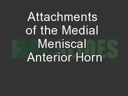

IntroductionPeripheral meniscal tears are located in the most vascular portion of the menisci and comprise 39Ð72 % [2, 3, 56, 69, 82] of all meniscal tears. % of all ACL tears [10, 53]. Conversely, the anterior horn of the medial meniscus is less commonly injured and, therefore, not well described in the literature. Chen et al. [ -cal techniques include open, outside-in, inside-out, and all-inside, in addition to nonoperative treatment in certain circumstances [4, 11, 14, 28]. Outcomes with these techniques have been favorable overall [3, 4, 20, 24, 28, 38, 40, 43, 46, 58 ) [54]. T
he literature reports the sensitivity and speciÞc-ity of joint line tenderness to be 55Ð85 % and 29.4Ð67 %, respectively [6, 33, 49, 85]. 7 Peripheral Meniscal Tears: How to ], and failure rates of meniscal repair dramatically increase with residual knee laxity [4, 23, 65]. Thus, knee laxity and meniscal tears should be addressed concurrently. These maneuvers will not be covered in this chapter but should be included for a thorough exam of the knee.As noted before, when all physical exam maneuvers and observations are used in combi-nation, the resulting diagnosis is more accurate th
an any test alone. Tenderness to palpation along the joint line is among the most common signs of meniscal tear, but joint effusion, crepitus, quadri-ceps atrophy, or lack of full knee range of motion (i.e., loss of extension more than 5¡) may also be noted on examination [ - age) are more common with the all-inside technique. Strke et al. [71] reported that regardless of the repair technique employed, there is a general trend of increasing failure rates with time (75Ð94 % of success in the Þrst year of surgery to 59Ð76 % beyond the fourth year). Of note, criteria for success and f
ailure approach is needed for this procedure. The sur-geon uses a spinal needle from an outside-in repair kit to pierce the overlying capsule. Transillumination of the skin can sometimes be useful to locate the tear and joint line when intro-ducing the needle. The spinal needle is then advanced through the superior or inferior side of the meniscus traversing the area of the tear. The inner cannula of the needle is removed, and a #1 PDS suture is placed through the needle into the joint. An arthroscopic grasper is used to secure the free end of the suture, while the needle is sub-se
quently removed, leaving the suture in the joint. A second pass is made with the spinal nee-dle through the corresponding side of the capsule in a similar manner as before. The inner cannula is again removed, and a looped suture retriever is passed through the second needle into the joint. The free end of the previously passed PDS suture is then placed through the looped retriever using a grasper, and the suture is pulled back out of the knee creating a mattress suture construct to secure the meniscal tear. Depending on the nature of the tear and surgeon preference, either a hori-zo
ntal or vertical mattress suture conÞguration can be utilized. Once the outside-in repair is complete, a minimal incision can be made with the knee ßexed to 90¡ where the exit of the suture is to be able to tie them in the surface of the cap-sule (Fig. 7.8).7.4.3 All-Inside TechniqueOnce the meniscal tear has been carefully assessed, the penetrating points of the meniscus are decided strategically. The meniscal depth probe is utilized at this point to determine the Fig. 7.8 Arthroscopic view of an anterior horn of a medial meniscus demonstrating PDS sutures penetrating the cap-sule
and the meniscus in a horizontal mattress conÞguration to repair the tear7 Peripheral Meniscal Tears: How to with these procedures [27, 51, 80, 84]. It is inherent that preserving the meniscus restores the joint congruity and loading, thus, preventing the development of osteoarthritis. Different techniques for repair have been described (all-inside, inside-out, outside-in, and trephination) for peripheral tears that allow for preservation of the meniscus. Repair of the meniscus improves clinical outcomes of pain, catching, and knee function using Tegner and Lysholm scores. Mean Lys
holm scores and Tegner scores for all-inside techniques are reported to be 90 and 6 respectively, while for the inside-out tech-nique, they are 88 and 5 respectively. When comparing the all-inside technique with the inside-out technique, no signiÞcant differences in clinical or anatomic failure rates (clinical failure, 11 range of motion (ROM) that is limited to the initial 2 weeks postoperatively. This early mobility facilitates postsurgical joint effusion resolution, normal range of motion restoration, and reduction of the scar formation. Passive ROM is completed with the patient
in the supine or seated position. Passive ROM is lim-ited to 0Ð90¡ during the Þrst 2 weeks and then progresses to full range of motion as tolerated by the patient. Isolated hamstring contraction is performed in the Þrst 6 weeks post-surgery to reduce meniscal stress through posterior tibial translation. Hyperextension of the tibio-femoral joint should be avoided at least for the Þrst 4 weeks in order to prevent stress on the meniscal repair. After this initial period of restriction, restoration of symmetrical exten-sion is encouraged for optimal tibiofemoral biomechanics. After 6 we
eks, if joint condi-tions and clinical examination deem appropri-ate, a progressive, weightbearing program is initiated. Also at this time, patients may begin the use of a stationary bike with low- resistance settings, and ! body weight leg presses to a maximum of 70¡ of knee ßexion. Starting 12 weeks postoperatively, additional increases in low-impact knee exercises may be permitted as tolerated. Patients are recommended to avoid deep squatting, sitting cross-legged, or performing any heavy lifting or squatting activities for a minimum of 4 months following surgery (Fig. 7.9). Con
clusionMeniscal tears constitute one of the most fre-quent pathologies in sports medicine. Due to the increasing understanding of its function and knee physiology, preservation of this tis-sue should be attempted in every case. A high index of suspicion is necessary at times to accurately diagnose some of these lesions, while meniscal tears are often evident in the physical exam and on imaging. Several tech-niques have been described with good to excellent reported outcomes. Determination of which technique to use depends on the ana-tomic meniscal region, the surgeonÕs prefer-ence,
and experience on each device. A robust rehabilitation protocol is mandatory to achieve the best results.7 Peripheral Meniscal Tears: How to -rior horn in the anterior cruciate ligament-deÞcient knee signiÞcantly inßuences anterior stability. Am J Bollen SR.Posteromedial meniscocapsular injury Meniscal ramp lesions: anatomy, incidence, diagnosis, and treatment. Orthopaedic J Sports Med. 2016;4 14. N, Corradi D, Garlaschi G, Zompatori M. Multidetector computed tomography arthrography of the knee: diagnostic accuracy and indications. Eur J Radiol. 2009;70:342Ð51. 17. De Flaviis L,
Scaglione P, Nessi R, Albisetti W. Ultrasound in degenerative cystic meniscal disease of the knee. Skeletal Radiol. 1990;19:441Ð5. 2002;179:1115Ð22. 42. cruciate ligament reconstruction patients. Arthroscopy. knee pain: diagnosis by the Þgure-4 test and treatment Arthroscopy. 2006;22:908.e901Ð4. 59. Petersen W, Tillmann B. Age-related blood and lymph supply of the knee menisci. A cadaver study. Acta Orthop Scand. 1995;66:308Ð12. 60. Rose NE, Gold SM. A comparison of accuracy between clinical examination and magnetic resonance imaging in the diagnosis of meniscal and anterior cru-
ciate ligament tears. Arthroscopy. 1996;12:398Ð405. 61. Rotterud JH, Sivertsen EA, Forssblad M, Engebretsen L, Aroen A. Effect of meniscal and focal cartilage lesions on patient-reported outcome after anterior cru-ciate ligament reconstruction: a nationwide cohort study from Norway and Sweden of 8476 patients with 2-year follow-up. Am J Sports Med. 2013;41:535Ð43. 62. Rutten MJ, Collins JM, van Kampen A, Jager GJ. Meniscal cysts: detection with high-resolution 2001;286:1610Ð20. 71. Meniscal 72. Staubli HU, Birrer S. The popliteus tendon and its fas-cicles at the popliteal hiatus:
gross anatomy and func-tional arthroscopic evaluation with and without anterior cruciate ligament deÞciency. Arthroscopy. 1990;6:209Ð20. 73. Steinbach LS. Calcium pyrophosphate dihydrate and calcium hydroxyapatite crystal deposition diseases: imaging perspectives. Radiol Clin North Am. 2004;42:185Ð205. vii 74. Westermann RW, Wright RW, Spindler KP, Huston LJ, Wolf BR. Meniscal repair with concurrent anterior cruciate ligament reconstruction: operative success and patient outcomes at 6-year follow-up. Am J Sports Med. 2014;42:2184Ð92. 84. Wroble RR, Henderson RC, Campion ER, el-Kho