
Neissereae Medical Microbiology By Asst Prof Dr Shaimaa Al Salihy Neisseriae Neisseriae The most important species are Neisseria gonorrhoea gonococci Neisseria meningitidis ID: 1039050
Download Presentation The PPT/PDF document "Lec . 7 Gram-negative cocci" is the property of its rightful owner. Permission is granted to download and print the materials on this web site for personal, non-commercial use only, and to display it on your personal computer provided you do not modify the materials and that you retain all copyright notices contained in the materials. By downloading content from our website, you accept the terms of this agreement.


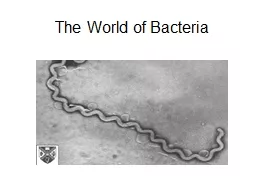
1. Lec. 7Gram-negative cocciNeissereaeMedical MicrobiologyBy:Asst. Prof. Dr. Shaima’a Al-Salihy
2. NeisseriaeNeisseriae:The most important species are:Neisseria gonorrhoea (gonococci)Neisseria meningitidis (meningococci) both of them are strict human pathogenfound inside polymprphnuclear cells. Others are normal flora of human respiratory tract.
3. Gram Negative cocciNeisseriaeMorphology:Gram negative cocci arranged in pairs.Kidney or coffee bean shape (the flat or concave sides are adjacent). Non-sporing, non-motileOxidase-positive that differentiate them from streptococci.Most are catalase-positive.
4. Gram Negative cocciNeisseriaeCulture:fastidious bacteria grow on enriched media; Mullar-Hinton, Modified Thayer-Martin or Heated blood agar (Chocolate agar). They produce convex glistening, non-pigmented, non-hemolytic colonies.They required 5% CO2 (Candle jar).Ferment sugars producing acid but not gas.
5. There are two pathogenic species for humans:Neisseria gonorrhoeae (Gonococci) GCNeisseria meningitidis (Meningococci) MCNon pathogenic e. g. Moraxella catarrhalis was previously named Branhamella catarrhalis (Neisseria catarrhalis).
6. Neisseria gonorrhoea:The causative agent of gonorrhea, ophthalmia neonatorum, and pelvic inflammatory disease (PID). Biotypes: - (T1, T2) virulent small brown colonies (piliated gonococci) posses pili which are adherence factors.- (T3, T4) a virulent large non pigmented colonies (no piliated gonococci). Virulence factors
7. Virulence factors:1.Pili : Pilin protein. Attachment to host cell and resist phagocytosis.2-IgA protease: split the IgA, major mucosal immunoglobulin.3.Cell wall components:Por: preventing phago-lysosome fusion Opa (Opacity): enhance adherence of gonococci within coloniesRmp (Reduction Modifiable protein)Lipooligosaccharide: possess endotoxic activity.Virulence factors
8. Pathogenesis:Gonorrhea:Transmitted by sexual contact.Gonoccocci attach themselves to the mucosal cells by pili and protein II (Opa)Invasion of cell.pass through the cells into the subepithelial space, thereby establishing the infection.Lipopolysaccharide: endotoxinIgA Protease: destroys immunoglobulin IgA
9. Clinical findings:In male:symptoms: urethra, purulent discharge, dysuriaincubation period:2-7 days after infectionComplication: epididymitis, prostatitis, periurethral abscessesIn female:symptoms: frequently asymptomatic. When symptomatic: cervicitis, vaginal discharge, dysuria, abdominal pain,Complication: ascending genital infection, salpingitis, tubo-ovarian abscess, pelvic inflammatory disease (PID)
10. Post-gonococcal urithritis (PGU): is common chronic sequelae of untreated or badly managed gonorrhoea, usually caused by different pathogens. Gonococcal ophthalmia neonatorum: An infection of the eye of newborn acquired during passage through infected birth canal.Disseminated gonococci infections (DGI) arthritis, skin lesion.
11. Lab. Diagnosis :- Specimens: pus, secretion from urethra, cervix, conjunctiva, throat and rectum. Gram stained smear: intracellular Gram-negative diplococci in PMNs (polymorphoneuclear cells) from urethral discharge, in men is sufficient for diagnosis while in women false positive because of the normal flora interference
12. Lab. Diagnosis :- 3. Culture: Chocolate agar. Thayer-Martin agar. (selective medium) which is chocolate agar containing antibiotics (vancomycin, colistin and nystatin to suppress the normal flora found in the specimen. Modified Thayer-Martin agar. same as(Thayer-Martin agar plus trimethoprim) The culture is incubated in an atmosphere containing 5% COZ (candle extinction jar) at 37°c. 4. Oxidase test positive. 5. Sugar fermentation the differentiation between GC and MC is made on the bases glucose and maltose. Ferment glucose only 6. Nucleic acid amplification test.
13.
14. Immunity: Antibody against Opa proteins and LOS.No protection against reinfection. due to the antigenic changes of the pili and outer membrane proteins.Treatment:PPNG have increased in prevalenceCefitriaxone, and Doxycycline for concomitant chlamydial infection substituted by erythromycin in pregnant women.
15. Neisseria meningitidis:The causative agent of meningitis, meningococcemia ( life-threatening form is Waterhouse-friderichsen syndrome ).Serogroups A, B, C, Y and W- 135. 5-30% of the normal population carrierVirulence factors: Polysaccharide capsule: antiphagocyticIgA proteaseLPS*Streptococcus pneumonia and Neisseria meningitides (more than 80% of cases bacterial meningitis in persons over 2 months of age).
16.
17. Pathogenesis and clinical findings: - portal of entry: nasopharynx and gain acsess to CNS:Invasion of blood stream: causing bacteremia.Direct contiguous spreadMeningitis is the most common complication of meningococcemia.Headache, vomiting and stiff neck.Waterhouse-Friderichsen syndrome: high fever, hemorrhagic rash, disseminated intravascular coagulation, and circulatory collapse
18.
19. Lab. Diagnosis :- 1. Specimens: blood, CSf and nasopharyngeal swap. 2. Direct-Gram stain smear of centrifuged CSF shows MC as typical Neisseria within polymorphonuclear leukocyte or extracellularly. 3. Culture: Choclate agar and incubated at 37oC with 5% CO2, or Thayer martin agar and modified TM. 4. Biochemical test :- --Oxidase test-positive. --Sugar fermentation test, ferment maltose and glucose. 5. Serology, antibodies to meningococcal polysaccharides can be measure by latex agglutination test or by measuring the bactericidal activity.
20. Immunity: - the immunizing antigen is capsular polysaccharideThere are 2 types of vaccine:Non conjuated tetravalent vaccine (Vaccine contains capsular polysaccharide of strains A, C, Y and W-135)Conjugated Treatment:- Penicillin G
21.
22.