
B enoit H ainaux E ric Lévêque N athalie C hemla P aris v C linic FRANCE Basics Of MRIHow I Do It AFIIM ISRA 2015 COIL Dedicated phased array Or flex coil ID: 525625
Download Presentation The PPT/PDF document "SHOULDER HOW I DO IT ?" is the property of its rightful owner. Permission is granted to download and print the materials on this web site for personal, non-commercial use only, and to display it on your personal computer provided you do not modify the materials and that you retain all copyright notices contained in the materials. By downloading content from our website, you accept the terms of this agreement.


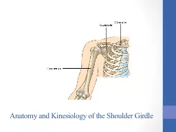
Slide1
SHOULDER HOW I DO IT ?
Benoit Hainaux, Eric LévêqueNathalie ChemlaParis v ClinicFRANCE
Basics Of MRI:How I Do It AFIIM -ISRA 2015Slide2
COIL
Dedicated phased arrayOr flex coil typeCloser to B0 center for a better FAT SATSlide3
COIL
What happens when coil touch the tunnel
T2 FAT SAT
T2 FAT SATSlide4
PATIENT POSITION
Lying on his back, opposite side rotated by 45°Arm along the body, neutral rotation, maintained with
velcro stripArm horizontalizedSlide5
SLICES POSITION
Coronal plane
Axial plane
Sagittal plane
“Y” shape of scapulaSlide6
CRITERIA OF SUCCESS
SSM
SSTSlide7
MAGIC ANGLE ARTIFACT
Appears on tendons, ligaments and cartilagesCollagen fibers are an anisotropic structure Normally hypo signal Slide8
MAGIC ANGLE ARTIFACT
If the tendon is 55°oriented compared to B0 axis Short TE sequences (SE T1 and PD, GE)It appears enhanced
B
0
55°
SE PD, TE=50ms
B
0
55°Slide9
MAGIC ANGLE ARTIFACT
Others anatomies and weightings…
SE T1 TE=7ms
SE T1 TE=12msSlide10
MAGIC ANGLE ARTIFACT
Others anatomies and weightings
SE PD TE=50ms
SE T1 TE=8msSlide11
MAGIC ANGLE ARTIFACT
To reduce this artifact :Change orientation of the tendonUse longer TET2 w sequences instead of PD wSE instead GE
SE PD TE=50ms
SE T2 TE=80ms Slide12
PROPELLER/BLADE/MULTIVANE
Radial sampling method of the k-spaceUsing rotating blades composed of phase-encoded linesMotion artefact reduction method
Without propeller
With propeller
PhaseSlide13
PROPELLER/BLADE/MULTIVANE
SE, GE, EPIOne blade is acquired in a single shotThen blade is rotated by a small angle (10-20°)This process continues till the entire k-space has been collectedSlide14
PROPELLER/BLADE/MULTIVANE
Center of k-space is oversampledRedundancy of information Data from each new blade can be compared to the previous oneData can be corrected Slide15
PROPELLER/BLADE/MULTIVANE
ExampleSlide16
PROPELLER/BLADE/MULTIVANE
ExampleSlide17
PROPELLER/BLADE/MULTIVANE
ExampleSlide18
Basics Of MRI:How I Do It AFIIM -ISRA 2015
FSET2 IN THREE ORTHOGONAL PLANS
NO FAT SAT DP AVOID MAGIC ANGLESlide19
FSE T1 ANATOMIC SEQUENCES
GOOD VISUALISATION OF BONE MARROW ANS MUSCLE TROPHICITYBasics Of MRI:How I Do It AFIIM -ISRA 2015
FSE T1
FSE T1
SUPRASPINATU
S
INFRASPINATUS
AND TERES MINOR
SUBSCAPULARIS
SUPRASPINATUSSlide20
Basics Of MRI:How I Do It AFIIM -ISRA 2015
MOTION ARTIFACT
FSE T1
FSE T2
GOOD CONTRAST BETWEEN FLUID AND MUSCULOSKELETASlide21
Basics Of MRI:How I Do It AFIIM -ISRA 2015
MR ARTHROGRAPHY
FSE T1
FSE T1 FAT SAT
FSE T2
3D FIESTASlide22
Basics Of MRI:How I Do It AFIIM -ISRA 2015
STERNO CLAVICULAR JOINT
FLEX COIL
PROPELLER TO AVOID MOTION AND CARDIAC ATIFACTS
T
1
T2Slide23
Basics Of MRI:How I Do It AFIIM -ISRA 2015
STERNO CLAVICULAR JOINTT1 AND T2 FSEPROPELLER TO AVOID MOTION AND CARDIAC ATIFACTS
T1
T2Slide24
AVOID MAGIC ANGLENO PD ON SHOULDER
Basics Of MRI:How I Do It AFIIM -ISRA 2015TAKE HOME MESSAGESlide25
Basics Of MRI:How I Do It AFIIM -ISRA 2015