
BCH 333 practical Lab 8 Objective To be familiar with Agarose gel electrophoresis principle Agarose gel electrophoresis is a method of gel electrophoresis used in biochemistry and molecular biology to separate and analyze DNA or RNA molecules by size ID: 917044
Download Presentation The PPT/PDF document "Agarose gel electrophoresis" is the property of its rightful owner. Permission is granted to download and print the materials on this web site for personal, non-commercial use only, and to display it on your personal computer provided you do not modify the materials and that you retain all copyright notices contained in the materials. By downloading content from our website, you accept the terms of this agreement.


Slide1
Agarose gel electrophoresis
BCH 333 [practical]
Lab# 8
Slide2Objective:
-To be familiar with Agarose gel electrophoresis principle.
Slide3Agarose gel electrophoresis:
is a method of gel electrophoresis used in biochemistry and molecular biology to separate and analyze DNA or RNA molecules by size.
-It is used mostly to determine the size and checking the purity of isolated DNA.
Principle:
-
Biomolecules [DNA or RNA] are separated by applying an electric field to move the negatively charged molecules [-] through an agarose matrix towards the anode [+] , and the biomolecules are separated by size in the agarose gel matrix.
-The largest molecules will have the most difficulty passing through the gel pores,
whereas the smallest molecules will move faster.
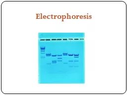
Slide4Separation of DNA molecules using agarose gel electrophoresis
Slide53- Agarose gel:
-Is a linear polymer of D-galactose and 3,6-anhydro-1-galactose and forms a gel that is held together by hydrogen bonds.
- Agarose is present in powder forms that are dissolved in buffer [TBE or TAE] close to boiling temperatures , then cooled down to around 40 degrees it sets and forms a gel . [polymerized]
-
The concentration of the material in the gel determines the size of the pores.
[high concentration of the gel
small pore size]
- Agarose gels have larger pore sizes compared to Dextran and polyacrylamide gels. This makes it useful for the analysis or separation of large globular proteins or long, linear molecules such as DNA.
Prepare agarose gel with 0.8%?
Slide6Slide7Buffer used:
-helps is deliver the electric current through the gel.
Buffer used is either TBE or TAE.
- TBE buffer:
is made with
Tris
/Boric acid/ EDTA.
- TAE buffer:
is made with
Tris
/Acetic acid/ EDTA.
Tracking Dye:
A dye such as
bromophenol
blue, Xylene
cyanol
or orange dye, is also included in the sample;
it makes it easier to see the sample that is being loaded and also acts as a marker of the electrophoresis front.
Note:
a glycerol or sucrose is included with the tracking dye , to render the samples denser than the running buffer (so that the
samples sink in
the well).
Slide8Slide9Slide10To load the sample,
we should take for example: 7 µl from the DNA sample + 2 µl from the dye
Slide11-After the samples of DNA are applied to the wells of agarose gel along with
the tracking dye.
-The power is turned on, after filling the buffer[?] in the tank to allows current to pass.
-The tracking dye and the DNA samples, are negatively charged [-] and migrate
towards the positive electrode [+].
-The DNA backbones contain negatively charged phosphate groups, which are attracted
to the positive electrode.
-Large DNA fragments retard earlier, while the smallest fragments move the fastest.
Slide12DNA: note the phosphate groups in the backbone are negatively charged.
Slide13Gel staining:
-The DNA in the gel needs to be stained and visualized, The reagent most widely used is the fluorescent dye
ethidium
bromide
"
EtBr
“
,
that emits orange light after binding to DNA.
Note: That the gel will be viewed under ultraviolet light.
[
Ethidium
bromide is a cyclic planar molecule that binds between the stacked base-pairs of DNA.].
Slide14Slide15http://www.beilstein-journals.org/bjoc/single/articleFullText.htm?publicId=1860-5397-10-312
Slide16"
EtBr“
orange
light after binding to DNA.
Slide17Determine the size of the DNA fragment:
-Since
agarose
gels separate DNA according to size, the size of a DNA fragment
may be determined from its electrophoretic mobility by running a number of standard
DNA markers of known sizes on the same gel.
Figure:
The 100
bp
DNA Ladder is suitable for sizing double-stranded DNA fragments from 100-2000
bp.
Slide18http://www.slideshare.net/hhalhaddad/forensic-lecture-6
Slide19Note that:
-The higher the voltage
the more quickly the gel runs.
[ Voltage
rate of migration].
-G
el concentrations must be chosen to suit the size range of the molecules to be separated.
Slide20SDS-PAGE
Vertical pace devices
Running buffer
Staining buffer [lamp]
De-staining buffer
Acrylamide gel
[stacking, separation gel]
Rf
value
Agarose
gel electrophoresis
Horizontal agarose devices
Running buffer
Stained and view using UV light
Agarose gel
Rf
value
Slide21http://learn.genetics.utah.edu/content/labs/gel/
https://www.youtube.com/watch?v=2UQIoYhOowM