/
CASE RECORD Acute bloody diarrhea with right sided colitis CASE RECORD Acute bloody diarrhea with right sided colitis

CASE RECORD Acute bloody diarrhea with right sided colitis - PDF document

christina
christina . @christina
Follow
342 views
Uploaded On 2022-09-07

CASE RECORD Acute bloody diarrhea with right sided colitis - PPT Presentation

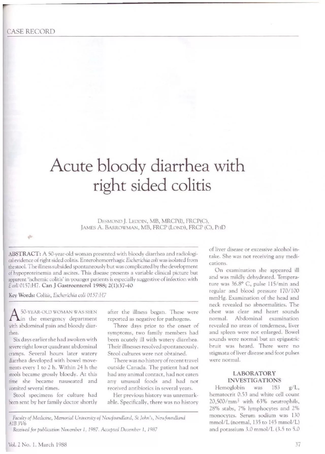
DESMOND J LEDDIN MB MRCPl FRCPC AMES A BARROWMAN MB FRCP LOND FRCP C Pl 10 ABSTRACT A SOyearold woman prescnced with bloody diarrhea and radiological evidence of righ c si ID: 952002

diarrhea coli disease colitis coli diarrhea colitis disease normal mmol illness days escherichia serum 0157 bloody watery colon reported

Share:

Link:

Embed:

Download Presentation from below link

Download Pdf The PPT/PDF document "CASE RECORD Acute bloody diarrhea with r..." is the property of its rightful owner. Permission is granted to download and print the materials on this web site for personal, non-commercial use only, and to display it on your personal computer provided you do not modify the materials and that you retain all copyright notices contained in the materials. By downloading content from our website, you accept the terms of this agreement.


Presentation Transcript

CASE RECORD Acute bloody diarrhea with right sided colitis DESMOND J. LEDDIN, MB, MRCP(l), FRCP(C), ]AMES A. BARROWMAN, MB, FRCP (LOND), FRCP (C), Pl 10 ABSTRACT: A SO-year-old woman prescnced with bloody diarrhea and radiologi­cal evidence of righ c sided col icis. Encerohemorrhagic Escherichia col, was isolated from the stool. The illness subsided spontaneously but was complicated by the development of hypoproteinemia and ascites. This disease presents a y s 2(1):37-40 Key Words: Colitis, fachenchia coli 0157:117 A50-YEAR·OLO WOMAN WAS SEEN in the emergency department with abdominal pain and bloody diar­rhea. Six days earlier she had awoken with severe right lower quadrant abdominal cramps. Several hours later watery diarrhea developed with bowel move­ments every I co 2 h. Within 24 h the stools became grossly bloody. At this time she became nauseated and vomited several rimes. Stool specimens for culture had been sent by her family doctor shortly after the illness began. These were reported as negative for pathogens. Three days prior to the onset 0f symptoms, rwo family members had been acutely ill with watery diarrhea. Their illnesses resolved spontaneously. Stool cultures were not obtained. There was no history of recent travel outside Canada. The patient had not had any animal contact, had not eaten any unusual foods and had not received antibiotics in several years. Her previous history was unremark­able. Specifically, there was no history Faculty of Medicine, Memorial University of Newfoundland, St John's, Newfoundland AIB3V6 Received for publication November I, 1987. Accepted December I, 1987 Vol. 2 No. I. March I Q88 of liver disease or excessive alcohol in­ take. She was not receiving any medi­cations. On examination she appeared ill and was mildly dehydrated. Tempera­ture was 36.8° C, pulse 115/min and regular and blood pressure 170/100 mm Hg. Examination of the head and neck revealed no abnormalities. The chest was clear and heart sounds normal. Abdominal examination revealed no areas of tenderness, lver and spleen were not enlarged. Bowel sounds were normal but an cpigastric bruit was heard. There were no stigmata of liver disease and foot pulses were normal. LABORATORY INVESTIGATIONS Hemoglobin was 183 g/L, hematocrit 0.53 and white cell count 20,500/mm3 with 63% neutrophils, 28% stabs, 7°0 lymphocytes and 2°0 monocytes. Serum sodium was l 30 mmol/L (normal, 135 to 145 mmol/L) and potassium 3.0 mmol/L (3.5 to 5.0 37 mmol/L). Serum chloride was 93 mmol/L (98 co l 10 mmol/L) and bi­carbonate 25.2 mmol/L (22 co 32 mmol/L). Blood urea was 4.8 mmol/L (3.0 ro 6.5 mmol/L) and crcatinine 72 µmol/L (40 co 130 µmol/L). Serum bilirubin was 4.8 µmol/L (less than 18 µmol/L), scrum asparrate aminotrans

­ferase (AST) was 17 iu/L (less than 38 iu/L), alanine aminotransferase (ALT) was I 7 iu/L (less ) a paLhogens. Sigmrndosc:opy revealed edematous colonic mucosa without ulceration. Following admission the patient ap­peared to deteriorate. She had fre- 4ucnc hlooc.ly bowel movements and c.lcvclopec.l f ia b left panel) anc.l this showeJ markec.l irregularity of the ascending and proximal transverse colon with thumb priming. This was consistent with the i.liagnosis of ischemic disease. The rest l l Six days after admission Escherichia co/10157:H7 positive for Verotoxin was isolated from the patient's stool. The patient was created with in­ Lravenous fluids and appropriate cor­rections were made to her electrolytes. The diarrhea setcled spontaneously. On the l 0th ay of her illness the white cell count was normal. By the I 2th day her howel frequency decreased to two bloodless formed stools per day. At this point she was noted clinically to have ascites with a fluid thrill. At this time serum albumin was 3 I g/L (normal 35 to 50 g/L) and urinaly­ sis was negative for protein. Serum hilirubin was 5 µmol/L, AST was 26 iu/L and ALT was 36 3 s anti partial thromboplastin time was 28 s y w showing rig he side of the colon, on ninth c.la) of illness (left hand pane/) and eighc week.1 lucer (righi hand panel) of her �illnesl at which time her bowel movemenci:. s es ( SLool cul cures, however, showed no growth of the common en­eerie pathogens. Colonic ischemia was considered and the finding of an epi­gascric bruit supported chis diagnosis. Sigmoidoscopc examination showed edematous mucosa bur no specific abnormalities. Barium enema showed changes in che right colon compatible with ischemic disease. Until relatively reccndy the diagno­sis would nor have been elucidated fur­ Lher. The isolation of cnrerohemor­rhagic E coli t chi a, pathogenic b acrribucablc co Bray and Beavan ( l). In che 1940s, in England, they recognized chaL y a frat:tion of childhood diarrheas were explicable on the basis of known pathogens. They postulated chaL E coli might, in some circumstances, be capable of produc­ing diarrhea. Rabbit antiserum was prepared to E coli cultured from the stool of a child with diarrhea. Using the antiserum they were able to de­monstrate the presence of che sa1rn: ) a cnterohemorrhag1 groups is clinically of little benefit be­cause pathogenesis is not yet clear and because che organism does not always e a members in the present case likely had enrero hemorrhagic E coli but yet had a CAN J GASTRt)cNTERL)L watery diarrhea without gross blood. The ability of emerohemorrhagic E coli co produce watery Jiarrhca has been reported previously (2). The O I 57:H7

strain of E coli was first recognized in lrish piglets in 1970 (3) as a cause of emeriris. Since then, ~poradic and clustered cases have been reported. Outbreaks have been described in homes for the ageJ (4) and in a day care centre (5). The disease 1s Lharacccn:cJ by an incubation pcrioJ of cl1rcc to nine Jays with a median of four days (4). The or­ganism may be ingested in LOntam1nat­ ed food. Hamburger from a restaurant chain was responsible for an outbreak in the UnircJ States (6). lnfouiun via fecal oral contamination and via fomites likely occun, (5). Typu.:ally the infection begins with nampy abdomi­nal pain followed by watery diarrhea which may become grossly bloody. There may be nausea anJ vommng hut fever 1s uncommon. Some patients have symptoms suggestivc of an upper respiratory infection (6). Laboratory evidence of tnflamma­non may be absent. The erythrocyte sedimentation rate and white cell count may be normal or only minimal­ ly elevated (6). Sigmoidoscop1c examination may be normal or show abnormaliues rang­ ing from edema co frank ulceranon (2). Colonoscopically the dbcasc may be most markeJ in the proximal colon and edema, inflammation and hcmor· rhage have been reported (2). Barium enema may be normal or show, as in chis case, changes suggestive of sub­ mucosal edema (2,6). The disease appears to show a predilection for the right colon and may mimic ischemic disease. Lt seems likely in milder cases, or in cases where chc patient is seen some time afccr the onset of the illness, that no s1gnifiLanc lesion may be seen e i is usually self-limited 11·1th a mean duration of eight days (2) but in the elderly and very young it may be fatal. A mortality rate of 16% was reported for an outbreak in Lon-Vol. 2 No. I. March 1988 don, Ontario 1n 1985 (4). There is an association with hemolytic uremic syn­drome a complication which carries a high mortality (2,5,7). The organism may be found in the scools for up to 14 days (8) but is more likely ro he detected if stools are col­lected within the first six days of illness (9). Treatment consists of fluid and elev trolytc replacement. Trimethoprim­sulfamethoxazole or ampic1llin may be effective in shortening the Juration and severity of illness (10). There arc, however, no controlled tnals of anti· hiotic efficacy. In the retrospective data presented hy Remis and col· leagues (2) the mean duration L)f illness in patients receiving amibiotics was 7. 5 days as compared to 8. 5 days in un­treated patients. Of particular impor· cancc would be a trial designed co show whee her antim1crob1al therapy rrevenced the development of disease and hemolytic uremiL syndrome in contacts. The organism

produces a toxrn 1denucal to chm of Shigelfo dvsenieriae type I ( 11 ). This toxin, designarcJ Verocoxin I, is cytotoxiL for HeLa and Vero cells (a cell line derived from monkey kidney cells). A second cytocoxin, Vcrocoxin 2, not neutral­ ized by antibody to the Shiga toxin may also be found. Some strains produce fimbriae which may be an important factor in determining v1rulcnce ( 12). Vcrotoxtn has been shown co cause platelet aggregation ( 13) anJ this may be important in the pathogenesis of the hemolytic uremic syndrome. E coli 0157:H? is not the only cmeric pathogen assoLiated with hemolyttL uremic syndrome. This associauon has also been reported for Salmonella, Shigella and Camp,lobaccer 1e11m1. ln the present case the disease fol­lowed a relatively benign course. There was no evidence of hemolys1s and renal E coli colitis fumnon remarncd normal. The dis­ ease subsided with suppon 1ve thcrap~. The case presented here is unusual as asc1tes has not previously been re­ported as a complicanon of the illness. The serum albumin was low possibly secondary to procein loss from the in­ flamed bowel. The low scrum onconc pressure due to hypoalbumincm1a no doubt contributed co chc asc1tes but docs not seem sufficcnt to explain it. There was no clrnical or laboratory l'Vi· dence of hepatic or pancreatic Jbcase and no evidence of periro111tis. When seen in follow-up eight weeks after the onset of the illness thl' paticrn was well with a normal serum albumin and no evidence of ascites. Repeat hart· um enema showed resolution of the colonic abnormalicies. E coli 0157:H? presents co the gas· troenrerologist in a variety of ways; as watery diarrhea, bloody diarrhl'a, colonic bleeding with no apparent cause or as apparent 1schcmit colitis. The reports of nght sided '1schcmic colius' m young adults (14) reporceJ 1n the liccraturc prior to recognition of this organism may be descriptions of cnterohcmorrhag1c E coli colitis. Microbiological laboratories should screen for this organism in all cases of bloody diarrhea when no ocher pathogen is found. Initial screening is based on the absent or slow fermenta­tion of sorbitol by rhb strain o( E coli. Over 90% of ocher strains of E coli fer­ ment sorbitol. If a nonfcrmeming strain is found it Lan be scrotyped to detcrmtnc i( 1r is cnterohcmorrhagic Ecoli. The gastroenrerologist should con­ sider E coli O 157:Hi as a cause of infec­ tious or apparent '1schemic' colitis when cultures are negative for the common pathogens. This 1s especially 11nporcanc if ban um enema or colono­ scopy shows disease predominantly af­ fecung the proximal colon. U'-1. 11::1\1\IE

l)J. 50 AN" ::,,'1::,, r !'RI :::,L:,,,TJ::I: A\'Et L NI: DJ,\RRI 11,1: 'i/\Nt,l./\l fl' et, a l'cxnmen baryte, unc colitc du cote: Jro1t. Les Loproculcurcs one rcvcle la prc~ence d'u nc souchc de E coli emcrohcmorrhagique. [I y cu unc arncltorat1on Jc la malaJ1e lCpendanc ellc fut compltqucc d'hypoproce111cm1e et J'ascice. La presentation dinique Je cette maladie est vanahlc mats c he:i lcs Jcuncs la dccouvcne d'unc 'colitc 1schcm1quc' suggcre fonrnc1u une infou1on u1uscc par E coli 0157 :H7. LED[)IN AND BARR~ 1\X'1'.tAN REFERENCES !.Bray J, Beavan TED. Slide agglutina­tion of Bacterium coli var. neapoliranum from summer diarrhea. J Pathol 1948; 60: 395-40 I. 2.Remis RS, McDonald KM, Riley LW, et al. Sporadic cases of hemorrhagic colitis associated with Escherichia coli OL57:H7. Ann Intern Med 1984; 101: 624-6. 3.Furowicz AJ, Orskov F. Two new Es­cherichia colr O antigens, 0150 and 0157, and one new K antigen, K92, in strains isolated from veterinary dis­eases. Acta Pacho! Microbial Scand (B) 1972; 80: 441-4. 4. Puduen D, Tuttle N, Korn D. cc al. Hemorrhagic colitis in a nursing home in Ontario. Can Med Assoc J 1986; 134: 50. *5. Spika JS, Parsons JE, Norden berg D, Wells JG, Gunn RA, Blake PA. Hemolyric uremic syndrome and diar­rhea associated with Escherichia coli 0157:H? in a day care center. J Pcdiacr I 986; 109: 287-91. *6. Riley LW, Remis RS, Helgerson SD, cc al. Hemorrhagic colitis associated with a rare Escherichia coli scrotype. N Engl J Med 1983; 308: 681-5. 7. Karmali MA, Petric M, Lim C, Flem­ing PC. Escherichia coli cycocoxin, haemolytic uremic syndrome, and haemorrhagic colitis. Lancet 1983; ii: 1299-1300. 8. Wells JG, Davis BR, Wachsmuth IK, et al. Laboratory investigation of hemor­rhagic colitis outbreaks associated with a rare Escherichia coli scrotype. J Clin Microbial 1983; 18: 512-20. 9.Pai CH, Gordon R, Sims HY, Bryan LE. Sporadic cases of hemorrhagic colitis associated with Escherichia coli 0157:H7. Ann Intern Med 1984; IOI: 738-42. JO. Levine MM. Antimicrobial therapy for infectious diarrhea. Rev Inf Dis 1986; (Suppl 2): S207-14. I I.O'Brien AD, Thompson MR, Caney JR, Formal SB. Production of a Sh11;ella dy~enceriae - like toxin by pathogcrnc Escherichia coli. Annual meeting of American Society of Microbiology 1977; No. BIOl. *12.Levinc MM. Eschenclt1a coli chat cause diarrhea: Enterotoxigcnic, entero­pathogcnic, cntcroinvasive, cnrero­hemorrhagic, and emcroadhcrcnt. J Inf Dis 1987; 155: 377-89. 13. Rose PE, Armour JA, Williams CE. Hill FGH. Verotoxin and neuramini­dase induced platelet aggregating ac­tivity in plasma: cheir possible role in the pathogenesis of che haemolytic uraemic J L 972; 4: 70-2. * Key reference Submit your manuscripts athttp://www.hindawi.