
Skeletal Muscle Tissue Cardiac Muscle Tissue Smooth Muscle Tissue Skeletal Muscle Tissue AKA Striated Muscles These are voluntary muscles Consciously controlled They are composed of distinctive light and dark bands ID: 920266
Download Presentation The PPT/PDF document "Muscular System Types of Muscles" is the property of its rightful owner. Permission is granted to download and print the materials on this web site for personal, non-commercial use only, and to display it on your personal computer provided you do not modify the materials and that you retain all copyright notices contained in the materials. By downloading content from our website, you accept the terms of this agreement.


Slide1
Muscular System
Slide2Types of Muscles:
Skeletal
Muscle
Tissue
Cardiac Muscle
Tissue
Smooth Muscle Tissue
Slide3Skeletal Muscle Tissue
AKA Striated Muscles:
These are voluntary muscles. Consciously controlled.
They are composed of distinctive light and dark bands
These muscles also have a limited capacity for regeneration due to the small number of cells that can divide.
Slide4Cardiac Muscle Tissue
Found only in the heart.
It is striated like skeletal muscle, however not as heavily.
It is not under conscious control. (Involuntary).
This type can regenerate under certain conditions,
Such as heart tissue transplants
Slide5Smooth Muscle Tissue
Non-striated
tissue and is found in the blood vessels, stomach, intestines, and airways.
This tissue is involuntary and has the greatest ability of any of the muscle tissue to regenerate. But it is still far more limited than other tissues.
Slide6Functions of the Muscular System
1. Movement
: Walking running and jogging are the movements that are voluntary and produced by the skeletal muscles along with the work of the nervous and skeletal system.
Slide72. Stabilizing Body Positions:
Skeletal Muscles are constantly contracting to keep your body posture upright. When any of these muscles are not used or become injured an problem in the skeleton may arise.
Slide83. Regulating Organ Volume:
Contractions of ring like sphincter muscles prevent overflow of organs such as the bladder or stomach.
4. Moving Substances within the Body
: Smooth muscles and cardiac Muscle do most of this work.
Cardiac Muscle pumps the blood though the blood vessels.
Smooth muscle help to regulate the diameter of the blood vessels to regulate blood flow. These muscles also are used in the gastrointestinal tract and the reproductive system.
Slide95. Producing Heat
: When a muscle contracts it produces heat. Shivering is an involuntary contraction of skeletal muscles that helps to warm the body.
Slide10Skeletal Muscle Tissue:
Fascia
: Fibrous band that surrounds muscles and other organs of the body.
Two types
1
. Superficial Fascia
2.
Deep Fascia
Slide111
. Superficial Fascia:
made up of fat and
areolar
connective tissue.
2.
Deep Fascia
: dense tissue and hold the muscles into together as well as separates them into functional groups.
Slide12Deep Fascia continued
Epimysium
wraps the entire muscle. This will include the
Tendons
. Tendons connect muscle to bone.
The
Perimyium
surrounds 10 to 100 muscle fibers called
fascicles
Endomysium
wraps individual muscle fibers.
Slide13Skeletal Muscles contract with the aid of nerves and blood vessels.
The
nerves
supply the electrical impulses and the
blood
delivers the necessary oxygen and nutrients needed for proper operation.
Slide14The oxygen and nutrients are required for the production of
ATP
,
Adenosine
Tri-phosphate
, that give the muscles the energy needed. The cells of the muscle have a large number of
Mitochondria
where ATP synthesis takes place.
Slide15Each skeletal muscle has an artery and a nerve that delivers the
nutrients and impulses.
They also have a few veins that carry waste products away.
The arteries branch out into small capillaries that supply each muscle fiber with the proper amount of materials.
Slide16Muscle Contraction and Function:
Proper muscle function is required for daily living and most of us have no idea of how a muscle works.
Slide17The muscle is made up of different components:
Muscle Fibers:
elongated cylindrical cells that run parallel to each other.
Sarcolemma
: a plasma membrane that covers each membrane.
Transverse Tubules
(T-Tubes). Tunnel like extensions of the
sarcolemma
.
Slide18Sarcoplasm
:
The cytoplasm of the muscle fiber. It contains large amounts of mitochondria that produce the ATP during contraction.
Sarcoplasmic
Reticulum:
A network of fluid filled tubules that contain calcium ions needed for contraction. These tubules run
thoughout
the
sarcoplasm
.
Myoglobin
:
Reddish pigment also found in the
sarcoplasm
that stores oxygen needed in ATP production.
Slide19Myofribriles
:
Cylindrical structures that extend along the entire length of the muscle fiber. It consists of two types of protein filaments.
Thin filaments
: composed of a protein called
Actin
b.
Thick Filaments
: Composed of a protein called
Myosin
Slide20Sacromeres
: The basic units of striated muscles and are created when the filaments overlap each other.
Z
-
discs
: these separate the
Sarcomeres
.
A
-
Band
: a darker area that extends the length of the
thick filament
. Found in the
sarcomere
.
Slide21H
-Zone: a narrow center area of the A-band that contains only thick filaments.
I
-Band: The lighter area on either side of the A-band that is made up of thin filaments.
The alternating darker A-bands and lighter I-bands give the
mucle
it striated appearance.
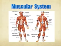
Slide22Muscles of the upper chest back and shoulder
Need to know these muscles
Deltoid
Latissimus
dorsi
Trapezius
Pectoralis
major
Pectoralis
minor
Slide23Trapezius
O: occipital bone, vertebrae
I: scapula
F: extend head, fix scapula
Slide24Deltoid
O: clavicle, scapula
I: humerus
F: abduct arm
Slide25Pectoralis Major
O: clavicle, sternum
I: humerus
F: flex, adduct arm
Slide26Pectoralis Minor
O: ribs
I: coracoid of scapula
F: depress scapula
Slide27Latissimus Dorsi
O: vertebrae
I: humerus
F: extend, adduct arm
Slide28Muscles of the Rotator Cuff
These are known as the SITS muscles
Supraspinatus
Infraspinatus
Subscapularis
Teres
minor
Slide29Supraspinatus
O: scapula
I: humerus
F: abducts arm, stabilize shoulder
Slide30Infraspinatus
O: scapula
I: humerus
F: laterally rotate arm
Slide31Subscapularis
O: scapula
I: humerus
F: medially rotate arm
Slide32Teres minor
F: laterally rotate, adduct arm
O: Lateral border of scapula
I: humerus
Slide33Muscles of the Upper and lower arm
The muscle that you need to know are the
Biceps
brachii
Triceps
brachii
Brachialis
Brachioradialis
Slide34Biceps brachii
O: scapula
I: radial tuberosity
F: flex forearm & arm
Slide35Triceps brachii
O: scapula & humerus
I: olecranon of ulna
F: extend forearm & arm
Slide36Brachialis
O: humerus
I: coronoid process of ulna
F: flex forearm
Slide37Brachioradialis
O: humerus
I: styloid of radius
F: flex forearm
Slide38Muscles of the Forearm
Need to know these muscles.
Slide39Supinator
O: humerus, ulna
I: radius
F: supinate forearm
Slide40Pronator teres
O: medial epicondyle of humerus, ulna
I: radius
F: pronate forearm
Slide41Palmaris longus
O: medial epicondyle of humerus
I: palmar aponeurosis
F: flex hand
Slide42Flexor carpi radialis
O: medial epicondyle of humerus
I: 2nd, 3rd metacarpals
F: flex, abduct hand
Slide43Flexor carpi ulnaris
O: medial epicondyle of humerus, ulna
I: carpals, 5th metacarpal
F: flex and adduct hand
Slide44Flexor digitorum superficialis
O: humerus, ulna, radius
I: middle phalanges 2-5
F: flex hand, phalanges
Slide45Flexor digitorum profundus
O: ulna
I: distal phalanges 2-5
F: flex hand, phalanges
Slide46Extensor carpi radialis longus
O: humerus
I: 2nd metacarpal
F: extend and abduct hand
Slide47Extensor carpi radialis brevis
O: lateral epicondyle of humerus
I: 3rd metacarpal
F: extend & abduct hand