
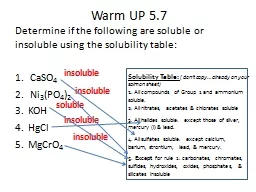
These insoluble proteins misfolded proteins are called amyloid More than 20 different proteins can aggregate to form the amyloid Amyloid is composed of nonbranching fibrils ID: 1007125
Download Presentation The PPT/PDF document "AMYLOIDOSIS Definition: A condition in ..." is the property of its rightful owner. Permission is granted to download and print the materials on this web site for personal, non-commercial use only, and to display it on your personal computer provided you do not modify the materials and that you retain all copyright notices contained in the materials. By downloading content from our website, you accept the terms of this agreement.


![BCH 302 [practical]](https://thumbs.docslides.com/578213/bch-302-practical-.jpg)


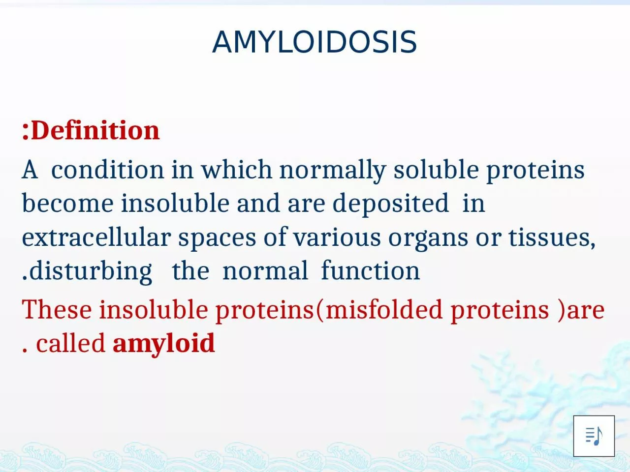
1. AMYLOIDOSISDefinition:A condition in which normally soluble proteins become insoluble and are deposited in extracellular spaces of various organs or tissues, disturbing the normal function.These insoluble proteins(misfolded proteins )are called amyloid .
2. More than 20 different proteins can aggregate to form the amyloid . Amyloid is composed of nonbranching fibrils, 7.5 to 10 nm in diameter, each formed of β-sheet polypeptide chains that are wound together.
3. The dye Congo red is commonly used to identify amyloid desposits in tissues. Congo red dye binds to the amyloid fibrils and produces a red-green birefringence.
4. Pathogenesis of Amyloid Deposition1- AL (amyloid light chain) protein Is produced by plasma cells and is made up of complete immunoglobulin (Ig) light chainsFor e.g. monoclonal B-cell proliferation (Plasma cell myeloma)
5. 2- AA (amyloid-associated) protein Is a protein derived from a larger serum precursor called serum amyloid-associated protein (SAA) that is synthesized in the in the liver.Is associated with chronic inflammatory disorders .
6. 3- Aβ amyloid (Amyloid beta) Aβ protein is derived from a much larger transmembrane glycoprotein called amyloid precursor protein (APP) by the action of proteolytic enzymes ; alfa & beta secretase. Aβ circulates in the plasma, CSF & brain interstitial fluid
7. Aβ is found in the cerebral lesions of Alzheimer disease. Plays a central role in the development of Alzheimer disease.
8. 4- Amyloid transthyretin (ATTR)(TTR)is a normal serum protein that binds & transport thyroxin and retinol.Mutations in the gene encoding transthyretin result in the production of a protein that aggregate and form amyloid deposits. The resultant diseases are called familial amyloid polyneuropathies.
9. 5- Aβ2- Microglobulin : affect patients on long term hemodialysis .A β2m is structurally similar to normal β2m . because it is not efficiently filtered through dialysis membrane and deposits in synovium , joints and tendon sheaths .
10.
11. Effects of AmyloidosisHistologically, the amyloid deposition is always extracellular and begins between cells, often closely adjacent to basement membranes. The clinical manifestations of amyloidosis depends on the organ involved
12. Kidney Amyloidosis of the kidney is the most common and most serious The amyloid deposits are found principally in the glomeruli
13. At first there is focal deposits within the mesangial matrix and diffuse or nodular thickenings of the basement membranes of the capillary loops. With progression , the deposition involves the capillary lumina and eventually leads to total obliteration of the vascular tuft.
14.
15.
16.
17. Liver Massive enlargement (as much as 9000 gm) Extremely pale, grayish, and waxy on both the external surface and the cut sectionHistologically , amyloid deposits first appear in the space of Disse (between hepatocytes ) and then progressively enlarge to encroach on the adjacent hepatic parenchyma and sinusoids .
18. The trapped liver cells undergo compression atrophy and are eventually replaced by sheets of amyloid*Normal liver function may be preserved even in the setting of severe involvement.
19. HeartAmyloid deposits are typically found throughout the myocardium , beginning Between myocardial fibers and eventually causing their pressure atrophy leading to cardiomyopathy with heart failure .
20.
21.
22.
23. Amyloidosis of the adrenals, thyroid, and pituitary is common but without apparent disturbance of function Nodular depositions in the tongue may produce macroglossia. CNS : Amyloid β protein associated with Alzheimer disease
24.