

introduction Between 300000 and 400000 surgical valve replacements are performed annually worldwide Aortic valve replacement accounts for the majority of surgical interventions followed by mitral and tricuspid valve replacement ID: 909106
Download Presentation The PPT/PDF document "Determinants of bioprosthetic aortic val..." is the property of its rightful owner. Permission is granted to download and print the materials on this web site for personal, non-commercial use only, and to display it on your personal computer provided you do not modify the materials and that you retain all copyright notices contained in the materials. By downloading content from our website, you accept the terms of this agreement.


Slide1
Determinants of bioprosthetic aortic valve degeneration
Slide2introduction
Between 300,000 and 400,000 surgical valve replacements are performed annually worldwide.
Aortic valve replacement accounts for the majority of surgical interventions, followed by mitral and tricuspid valve replacement.
Slide3Introduction
ACC guidelines
<50 years: mechanical aortic valves
>50, <70 years: either
>70: bioprosthetic aortic valves
ESC Guidelines
<60 years: mechanical aortic valves
>60, <65: either
>65: bioprosthetic aortic valves
Slide4introduction
Recently, there has been a trend towards increased use of biological valves in younger patients.
One justification for this approach is the possibility of transcatheter valve-in-valve replacement in case of valvular degeneration.
In addition, recent observational studies showed that in patients 50 to 69 years of age, mortality was not related to the choice of mechanical or biological aortic or mitral valve material.
Slide5introduction
Structural valve degeneration remains the major determinant of bioprosthetic valve durability.
Previous studies defined bioprosthetic valve degeneration as the need for reoperation, because careful and regular echo follow-up was not available.
More recently, bioprosthetic valve degeneration has been defined according to echocardiographic criteria.
On the basis of changes of
transprosthetic
gradients and severity of regurgitation, the term “valve
haeodynamic deterioration” (VHD) has been introduced.
Slide6Aim of this study
Was to assess the incidence and
mdoe
of VHD, as well as associated factors.
Slide7methods
Between 1994 and 2009, 504 consecutive adult patients undergoing AVR, mitral valve replacement, or tricuspid valve replacement with bioprosthetic heart valves agreed to participate in the present observational study at the Vienna General Hospital.
Mitral and tricuspid valves were excluded from this study due to the small numbers (MVR: 36, TVR: 2).
From study entry, all data were collected prospectively.
Slide8definitions
Clinically relevant VHD: elevated mean
transprosthetic
gradient (
>
30mmHg) and/or at least moderate
intraprosthetic regurgitation.Subclinical VHD: elevated mean transprosthetic gradient (>
20mmHg) and/or at least mild to moderate intraprosthetic regurgitation.VHD: presence of clinically relevant VHDPatient-prosthesis mismatch
(PPM): effective orifice area indexed to body surface are of
<
0.8cm
2
/m
2
.
Slide9Clinical measures and follow-up
Clinical evaluation, medical history, blood samples, and a TTE were collected at baseline.
Traditional CV risk factors were recorded according to the respective guidelines, and the European System for Cardiac Operative Risk Evaluation score was calculated.
Patients were followed up before discharge from the hospital, after 6 months, and every 1 to 2 years thereafter.
Follow up comprised of clinical evaluation, electrocardiography, TTE, and laboratory assessment.
Primary study endpoint: All-cause mortality.
Slide10Echocardiographic assessment
Cardiac morphology was assessed using diameter in standard 4- and 2-chamber views.
LVEF was calculated using the biplane Simpson method.
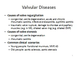
Valvular stenosis and regurgitation were quantified using an integrated approach and were graded as none, mild, mild to moderate, moderate, moderate to severe, or severe.
Systolic pulmonary artery pressure was calculated by adding the peak TR systolic gradient to the estimated CVP.
Slide11Echocardiographic assessment
To determine delay to VHD development, the date of first detection of VHD was included in the final analysis.
Annualised
change in mean gradient (mmHg/year) was calculated by dividing the difference between the latest and first follow-up echo by the time interval between the 2 assessments.
VHD incidence per year: number of VHD patients divided by the total number of years with echo follow up.
Slide12results
466 patients underwent surgical bioprosthetic AVR
The median follow-up period was 113.2 months
4,023 valve-years.
Echocardiographic and laboratory follow-up was complete in 82.2% of patients (n = 383)
Slide13Slide1430-40
29-45
39-59
20-33
Slide15Factors related to the occurrence of VHD
70 patients (18.3%, 4.8 per valve-year) developed VHD during a median follow-up of 33 months.
Modes of VHD:
Stenosis: 45 patients
Regurgitation: 16
Both: 9.
Median delay from surgery to VHD: 32.4 months.
Slide16Factors related to the occurrence of VHD
Cutoff value of 2.1mg/dL (185.6
umol
/L) serum creatinine was found to be closely associated with VHD development.
Further parameters associated with VHD on univariate and multivariate analysis were:
Porcine tissue prosthesis
Arterial hypertension
PPM
Slide17Impact of age on the time course of VHD
Elderly subjects showed shorter event-free survival with regard to the development of VHD
<70 years: 133.5 months
70-80: 129.1 months
>80 years: 100.3 months
VHD rates:
<70: 2% per valve-year
70-80: 4.7% per valve-year
>80: 5.4% per valve-year
Median delay from subclinical to clinically relevant VHD was 24.2 months.
Slide18Factors associated with outcomes
After a median follow up period of 113.2 months, 69.3% of patients had died (323).
CV death: 59.4% (192)
Slide19discussion
Whether to implant a mechanical or a bioprosthetic valve, particularly in patients 50-70 years of age is a matter of discussion.
Risks associated with the need for AC after mechanical valve implantation have recently led to an increasing use of bioprosthetic valves in patients older than 50 years.
Trend has been supported by clinical data, suggesting extended durability of new-generation
bioprosthesis
.
Percutaneous valve-in-valve tech provides a new, less invasive option to treat potential degeneration of bioprosthetic valves.
Slide20discussion
Present study is the first to use thorough echocardiographic follow-up at a single echo lab to provide long-term data on VHD-associated factors, including age.
Data collected at baseline and follow up.
Followed up for a medial of 9.4 years.
Incidence of clinically relevant VHD of 18.3% (n=70, 4.8% per valve-year)
Median delay to VHD: 32.4 months
Event-free survival with regard to development of VHD was significantly shorted in elderly subjects.
Risk factors for the onset of VHD: ↑ creatinine, porcine tissue valves, arterial hypertension, PPM.
Slide21Discussion – RF for Bioprosthetic VHD
Risk factors previously reported to be associated with bioprosthetic degeneration include young age, mitral valve position, end-stage renal disease, HTN, metabolic syndrome, lipid-mediated inflammation, diabetes, and PPM.
Data on porcine valves are conflicting.
In this study, VHD was associated with porcine valves, renal impairment, HTN and PPM, but young age was not a factor.
Strong trend toward shorter time to VHD in patients older than 80 years.
However, this group only had 64 patients, so it should be interpreted with caution.
Slide22Discussion – RF for Bioprosthetic VHD
Valvular calcification has previously been proposed as a major mechanism of
bioprosthesis
denegeration
.
To avoid immune system reaction against xenograft antigens, bioprosthetic leaflet tissue is fixed in glutaraldehyde, there rendering the bioprosthesis “immunologically inert”.However, residual
xenoantigents on pre-treated valves may induce immune response and cause valvular calcification.
Slide23Discussion – RF for Bioprosthetic VHD
Porcine and bovine tissue exhibit different set of antigens, presumably making tissue less or more prone to immunoreactivity.
Might provide an explanation for the increased incidence of VHD in porcine valves in this study.
Free aldehyde groups in pre-treated prosthesis bind calcium ions and phospholipids, resulting in accelerated mineralization in states of hypercalcemia and hyperphosphatemia.
Renal impairment frequent
causesly
dysregulation of
phosphocalcic metabolism, thereby leading to a more rapid progression of valvular degeneration.
Slide24Discussion – RF for Bioprosthetic VHD
Patients with renal impairment have a high
coprevalence
of arterial hypertension, which may additionally accelerate VHD by increasing diastolic closure stress on the prosthesis.
Another factors is PPM.
Elevated
transprosthetic gradients causes mechanical leaflet stress.over time, leaflet thickening and calcification may ensue, making PPM a substantial factor for valvular degeneration.
Slide25Discussion – RF for Bioprosthetic VHD
Age as a determinant of VHD has been investigated in a limited number of studies.
However, a number of these studies were retrospective in design, and echo assessment was not
standarised
.
Younger age at the time of valve surgery has been reported to be a risk factor for reoperation, by other studies.
However, they lacked systemic, prospective echo follow-up or did not provide data on echocardiography at all.
Time to the development of valvular degeneration was defined as freedom from reoperation.Reluctance to perform reoperation in elderly patients and incomplete echo assessment might thus have caused substantial bias.
Slide26Outcomes after SAVR
Few data on determinants of survival after surgical implantation of
bioprosthesis
.
Across most studies, mechanical and bioprosthetic valves were not
analysed
separately, thus hindering proper comparison with this study’s data.Renal impairment has been reported to inversely affect both in-hospital and long-term outcomes after valve replacement therapy.
Similar finding in the current study.
Slide27Outcomes after SAVR
Patients with diabetes are not only known to be more likely to develop cardiovascular disease, but they are further disadvantaged with worse outcomes after cardiac surgery.
Furthermore, concomitant CABG proved to have a significant impact on all-cause mortality in previous studies.
Haemodynamic
deterioration of aortic
bioprosthesis
has recently been established as an important predictor of outcomes.
Slide28limitations
Data collected in a single center setting.
Center-specific bias cannot be excluded.
Use of various valve types with different
haemodynamic
profiles limits the interpretation of data.
Group of patients >80 years old was small and follow-up was shorter when compared to other groups.Echo follow-up was incomplete in 17.8% of patients (83 patients), potentially creating a selection bias in the population eligible for final analysis.
Slide29conclusions
On the basis of echo data, the authors observed a significant incidence of VHD in bioprosthetic aortic valves.
VHD was associated with renal impairment, the use of porcine tissue valves, arterial hypertension, and PPM.
Patients younger than 70 years were not affected by
faster VHD.