
Lec 2 2 nd Grade Fall Semester 20212022 Goran Noori Saleh MSc At HMUgorannori tiueduiq 24102021 CLASSIFICATION OF EPITHELIA Simple epithelium Single layer of cells resting on a basement membrane ID: 904741
Download The PPT/PDF document "Epithelial tissue Histology and Histopat..." is the property of its rightful owner. Permission is granted to download and print the materials on this web site for personal, non-commercial use only, and to display it on your personal computer provided you do not modify the materials and that you retain all copyright notices contained in the materials. By downloading content from our website, you accept the terms of this agreement.


Slide1
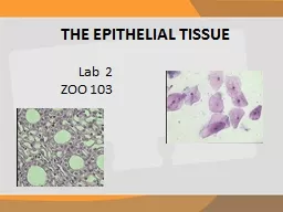
Epithelial tissue
Histology and Histopathology
Lec. 2
2nd Grade – Fall Semester 2021-2022
Goran Noori Saleh
MSc. At HMU(goran.nori@tiu.edu.iq)
24/10/2021
Slide2CLASSIFICATION OF EPITHELIA
Simple epithelium: Single layer of cells resting on a basement membrane.
squamous epithelium.cuboidal epithelium.columnar epithelium.Stratified epithelium
: Epithelia which consist of multiple layers with the basal layer resting on the basement membrane. The epithelium is named according to the shape of cells of the most superficial layer. Stratified squamous:Stratified cuboidal:Stratified columnar:
Pseudostratified epithelium: In true sense this is a simple epithelium as each cell rests on the basement membrane. This epithelium gives an appearance of a multilayered epithelium due to unequal height and shape of cells.Transitional epithelium:
In this type of multilayered epithelium all layers are made up of cuboidal, polygonal, or round cells. The cells toward the surface of the epithelium are round. As transitional epithelium is confined to the urinary tract, it is also called urothelium.
Slide3CLASSIFICATION OF EPITHELIA
Stratified epithelium:Stratified squamous:
DescriptionThis type of epithelium is made up of several layers of cells The cells of the deepest (or basal) layer rest on the basement membrane, they are usually columnar in shape. Lying over the columnar cells there are polyhedral or cuboidal cells. As we pass toward the surface of the epithelium these cells become progressively more flat, so that the most superficial cells consist of flattened squamous cells.
The nuclei are oval in basal layer, rounded in the middle layer, and transversely elongated in the superficial layers, the surface layer shows squamous cells with the flattened nuclei.Stratified squamous epithelium can be:non-keratinized: In situations where the surface of the squamous epithelium remains moist, the most superficial cells are living and nuclei can be seen in them.
Keratinized: At places where the epithelial surface is dry (as in the skin) the most superficial cells die and lose their nuclei. These cells contain a substance called keratin, which forms a non-living covering over the epithelium.
Slide4CLASSIFICATION OF EPITHELIA
Stratified epithelium:Stratified squamous:
Stratified squamous epithelium (both keratinized and non-keratinized) is found over those surfaces of the body that are subject to friction. As a result of friction the most superficial layers are constantly being removed and are replaced by proliferation of cells from the basal layer. This layer, therefore, shows frequent
mitoses.LocationKeratinized stratified squamous epithelium covers the skin of whole of the body and forms the epidermis. Non-keratinized stratified squamous epithelium covers wet surfaces exposed to wear and tear. It is seen lining the mouth, the tongue, the oro
- and laryngopharynx, the esophagus, the vagina, and the cornea. Under pathological conditions the epithelium in any of these situations may become keratinized. Function
It is protective in nature. Keratin prevents dehydration of underlying tissue.
Slide5CLASSIFICATION OF EPITHELIA
Stratified epithelium:Stratified Columnar or c) Cuboidal Epithelium
DescriptionThis epithelium consists of two or more layers of columnar or cuboidal cells.
Location Stratified cuboidal and columnar epithelium is seen in large ducts of exocrine glands like sweat glands, pancreas, and salivary glands. Function
Like all stratified epithelia it is protective in function and it also helps in conducting the secretion of the glands.
Slide6CLASSIFICATION OF EPITHELIA
Pseudostratified epithelium Description
It is not a true stratified epithelium but appears to be stratified. Normally, in columnar epithelium the nuclei lie in a row, toward the basal part of the cells. Sometimes, however, the nuclei appear to be arranged in two or more layers giving the impression that the epithelium is more than one cell thick. The cells are attached to the basement membrane but are of different heights, some cells are short and basal, while others are tall and columnar.The epithelium may bear cilia (ciliated epithelium) and may contain goblet cells. The cilia are capable of movement. At some sites this epithelium may display stereocilia
.
Slide7CLASSIFICATION OF EPITHELIA
Pseudostratified epithelium Location
Non-ciliated pseudostratified columnar epithelium is found in some parts of the auditory tube, the ductus deferens, and the male urethra (membranous and penile parts). Ciliated pseudostratified columnar epithelium is seen in the trachea and in large bronchi.Pseudostratified columnar epithelium with stereocilia
(long microvilli) is seen in epididymis.Function
The tall columnar cells are secretory in nature.while the short, basal cells are stem cells which constantly replace the tall cells. The cilia help in clearance of the mucous. The
stereocilia help in absorption.
Slide8CLASSIFICATION OF EPITHELIA
Transitional epithelium: In this type of multilayered epithelium all layers are made up of cuboidal, polygonal, or round cells. The cells toward the surface of the epithelium are round. As transitional epithelium is confined to the urinary tract, it is also called
urothelium.DescriptionThis is a multilayered epithelium and is 4 to 6 cells thick. It differs from stratified squamous epithelium in that the cells at the surface are not squamous. The deepest cells are columnar or cuboidal. The middle layers are made up of polyhedral or pear-shaped cells. The cells of the surface layer are large and often shaped like an
umbrella. In the urinary bladder, it is seen that cells of transitional epithelium can be stretched considerably without losing their integrity. When stretched it appears to be thinner and the cells become flattened.
Slide9CLASSIFICATION OF EPITHELIA
Transitional epitheliumLocation
Transitional epithelium is found in the renal pelvis and calyces, the ureter, the urinary bladder, and part of the urethra. Because of this distribution it is also called urothelium.
Function At the surface of the epithelium the plasma membranes are unusual. Embedded in the lipid layer of the membranes there are special glycoproteins. It is believed that these glycoproteins make the membrane impervious and resistant to the toxic effects of substances present in urine, and thus afford protection to adjacent tissues.
Slide10Histopathology (Medical correlation)
In individuals with chronic vitamin A deficiency, epithelial tissues of the type found in the bronchi and urinary bladder
may gradually be replaced by stratified squamous epithelium.The
ciliated pseudostratified epithelium lining the bronchi of smokers can also be transformed into stratified squamous epithelium by metaplasia
.Some epithelial cells are prone to abnormal growth or dysplasia, which can progress to precancerous growth called
neoplasia. Early neoplastic growth is often reversible and does not always result in cancer. Under certain abnormal conditions, one type of epithelial tissue may undergo transformation into another type in another reversible process called metaplasia.
Homework:
what we mean by
Dysplasia
Slide11BASEMENT MEMBRANE
Epithelial cells rest on a thin basement membrane
. In multi-layered epithelia, the deepest cells lie on this membrane. A distinct basement membrane cannot be seen in hematoxylin and eosin (H & E) preparations, but can be well demonstrated using the periodic acid Schiff (PAS) method. Under the EM a basement membrane is seen to have a basal lamina (nearest the epithelial cells) and a reticular lamina or
fibroreticular lamina (consisting of reticular tissue and merging into surrounding connective tissue). The basal lamina is divisible into the lamina densa containing fibrils; and the lamina lucida which appears to be transparent. The lamina
lucida lies against the cell membranes of epithelial cells.
Slide12BASEMENT MEMBRANE
Functions of Basement Membrane It provides adhesion on one side to epithelial cells (or parenchyma); and on the other side to connective tissue.
It acts as a barrier to the diffusion of molecules. May play a role in cell organization, as molecules within the membrane interact with receptors on cell surfaces.
Slide13PROJECTIONS FROM THE CELL SURFACE
Cilia
These can be seen with the light microscope, as minute hair-like projections from the free surfaces of some epithelial cells. In the living animal cilia can be seen to be motile. Functions of CiliaMovements of cilia lining the respiratory epithelium help to move secretions in the trachea and bronchi toward the pharynx.
Ciliary action helps in the movement of ova through the uterine tube, and of spermatozoa through the male genital tract. Cilia-like structures perform a sensory function. They may be non-motile, but can be bent by external influences. Such “cilia” present on the cells in the olfactory mucosa of the nose are called olfactory cilia, they are receptors for smell. Similar structures called kinocilia are present in some parts of the internal ear.
Slide14PROJECTIONS FROM THE CELL SURFACE
Microvilli
: are finger-like projections from the cell surface that can be seen by electron microscope. In some cells the microvilli are not arranged so regularly. With the light microscope the microvilli of such cells give the appearance of a brush border.Function of Microvilli
Microvilli are non-motile processes which greatly increase the surface area of the cell and are, therefore, seen most typically at sites of active absorption, e.g. the intestine, and the proximal convoluted tubules of the kidney.
Slide15Histopathology (Medical correlation)
Celiac disease, also called gluten-sensitive enteropathy or sprue, is a disorder of the small intestine in which one of the first pathologic changes is
loss of the microvilli brush border of the absorptive cells. This is caused by an immune reaction against the wheat protein gluten during its digestion, which produces diffuse enteritis (intestinal inflammation), changes to the epithelial cells leading to malabsorption, and eventually to pathologic changes in the intestinal wall. The malabsorption problems and structural changes are reversible when gluten is removed from the diet.
Slide16PROJECTIONS FROM THE CELL SURFACE
3.
Stereocilia are very long, thick microvilli . They are non-motile. Stereocilia are seen on receptor cells in the internal ear, and on the epithelium of the epididymis.
Function of StereociliaThey increase the cells surface area for absorption in epididymissignal generation in hair cells of internal ear.
Slide17